Review
. 2011 Dec 28;11(1):209-23.
doi: 10.1102/1470-7330.2011.0030.

Thyroid nodules: risk stratification for malignancy with ultrasound and guided biopsy

Affiliations
Review

Thyroid nodules: risk stratification for malignancy with ultrasound and guided biopsy

Gopinathan Anil et al. Cancer Imaging. .

Abstract

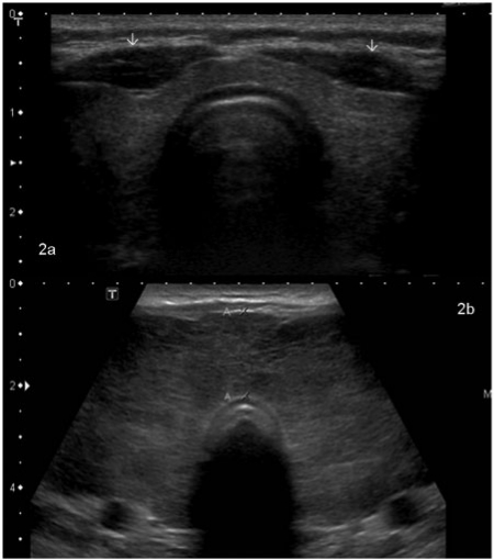
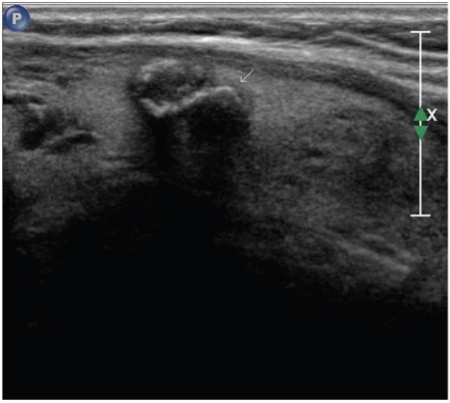
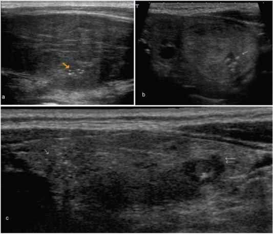
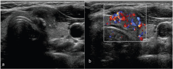
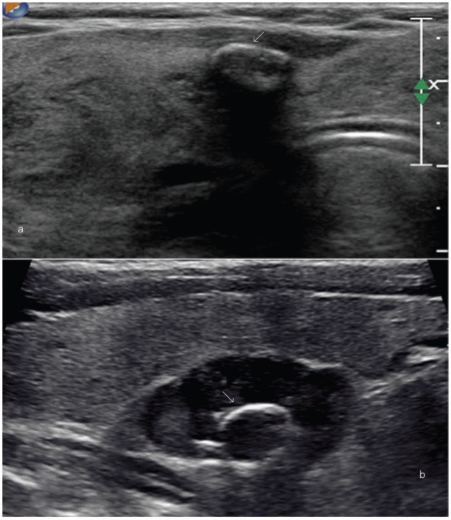
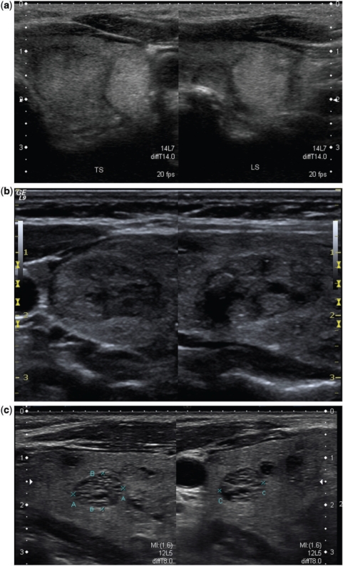
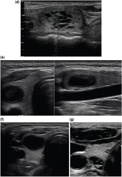
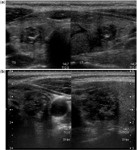
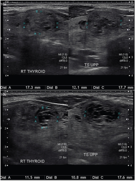
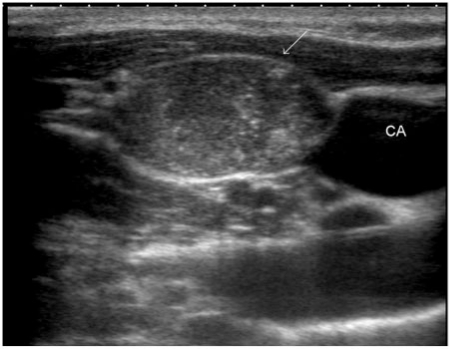
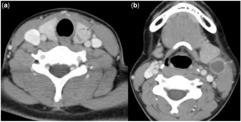
Replacing palpating fingers with an ultrasound (US) probe has resulted in an epidemic of thyroid nodules. Despite the high prevalence of thyroid nodules in the general population, thyroid malignancy is rare. Although no imaging modality can accurately predict the nature of every nodule, high-resolution US is the most sensitive, easily available and cost-effective diagnostic test available to detect thyroid nodules, measure their dimensions and identify their structure. The presence of calcifications, irregular spiculated outline, hypoechogenicity in a solid nodule, chaotic intranodular vascularity and an elongated shape are well-known US features of malignancy in thyroid nodules. Cervical lymph node metastasis and extrathyroidal extension of a thyroid nodule are highly specific for malignancy but seen infrequently. Spongiform nodules, purely or predominantly cystic nodules, nodules with well-defined hypoechoic halo and echogenic as well as isoechoic nodules are usually benign. None of the US characteristics have 100% accuracy in detecting or excluding malignancy. Fine-needle biopsy is currently the best triage test for pre-operative evaluation of a thyroid nodule. There is no significant difference in the risk for malignancy between palpable and non-palpable nodules and size is not a reliable indicator for their malignant potential. The best tool for risk stratification for malignancy in thyroid nodules is US and guided biopsy of nodules with suspicious imaging features. This is especially relevant in patients with multinodular goitre.


Figures

Figure 1.
Figure 1.
Any lesion within the thyroid gland that is radiologically distinct from the surrounding thyroid parenchyma qualifies as a thyroid nodule. (a) A large nodule (arrow) in the right lobe that was palpable at clinical examination. High-resolution ultrasound can detect much smaller nodules. (b,c) Longitudinal and axial images of the same lesion (shown by arrows) that measures 1×2 mm in size.
Figure 2.
Figure 2.
Transverse grayscale mode image of a normal thyroid gland that is uniformly echogenic relative to overlying strap musculature (arrows) (a). In comparison, (b) shows a thyroid gland with heterogeneous parenchymal echotexture and diffuse enlargement consistent with thyroiditis. This patient had Graves disease.
Figure 3.
Figure 3.
This grayscale image of the left lobe of the thyroid along its long axis in a patient with multinodular goitre shows a nodule with coarse chunky calcification (arrow). At histology, this was found to be benign. However, coarse calcifications when present in a solitary nodule are highly suspicious for malignancy.
Figure 4.
Figure 4.
These images show microcalcifications in various thyroid pathologies. Although one of the most specific US features of malignancy, microcalcifications may also be seen in benign conditions. (a) An enlarged heterogeneous thyroid gland with biopsy proven benign microcalcifications (arrow), in this patient with Hashimoto thyroiditis. (b) The long axis view of a well-defined, isoechoic left lobe thyroid nodule with hypoechoic rim shows a cluster of microcalcifications (arrow). At histology, this was diagnosed as a follicular adenoma. (c) The long axis view of the right lobe of the thyroid shows an ill-defined hypoechoic area with several microcalcifications at the cranial third (single arrow) and an irregular hypoechoic nodule with few microcalcifications at the lower third (double arrow). Surgery confirmed multifocal papillary thyroid carcinoma.
Figure 5.
Figure 5.
An invasive papillary carcinoma. (a) The transverse grayscale image shows a hypoechoic nodule with irregular outlines infiltrating the overlying strap muscle. Both coarse and microcalcifications are seen in this nodule. At colour Doppler interrogation (b) there is chaotic vascularity in the entire nodule.
Figure 6.
Figure 6.
Examples of eggshell calcifications. (a) A bulky thyroid gland in a patient with multinodular goitre shows a small nodule with partial rim calcification (arrow). (b) There is a well-defined hypoechoic solid nodule with eggshell calcification (arrow) within. This was a follicular adenoma at surgical excision.
Figure 7.
Figure 7.
Various histology proven benign thyroid nodules. (a) An isoechoic and an echogenic nodule. The isoechoic nodule is seen better in the transverse section (TS) and the echogenic one in the longitudinal section (LS). Besides the echotexture that favours benignity in both these nodules, they also show a well-defined hypoechoic halo that strongly favours benignity. (b) This mixed echotexture solid nodule shows a discrete hypoechoic rim along its entire circumference. A complete uniform halo around a nodule is highly suggestive of benignity, with a specificity of 95%. (c) The well-defined nodule here shows an aggregation of innumerable tiny cystic spaces giving it a spongiform appearance. Such nodules are usually benign and do not need biopsy. (d) Here the isoechoic nodule has multiple cystic spaces occupying more than two-thirds of its volume with a well-defined hypoechoic halo. Such microcystic components in more than 50% of the nodule volume are 99.7% specific for identification of a benign nodule[6]. (e,f) Predominantly cystic and purely cystic thyroid nodules. Thyroid cancer is not common in predominantly cystic nodules and purely cystic nodules are almost never malignant. (g) This transverse image of the right thyroid lobe shows a cyst with multiple echogenic foci with posterior ring-down artefacts. This appearance is diagnostic of a colloid cyst.
Figure 7.
Figure 7.
Various histology proven benign thyroid nodules. (a) An isoechoic and an echogenic nodule. The isoechoic nodule is seen better in the transverse section (TS) and the echogenic one in the longitudinal section (LS). Besides the echotexture that favours benignity in both these nodules, they also show a well-defined hypoechoic halo that strongly favours benignity. (b) This mixed echotexture solid nodule shows a discrete hypoechoic rim along its entire circumference. A complete uniform halo around a nodule is highly suggestive of benignity, with a specificity of 95%. (c) The well-defined nodule here shows an aggregation of innumerable tiny cystic spaces giving it a spongiform appearance. Such nodules are usually benign and do not need biopsy. (d) Here the isoechoic nodule has multiple cystic spaces occupying more than two-thirds of its volume with a well-defined hypoechoic halo. Such microcystic components in more than 50% of the nodule volume are 99.7% specific for identification of a benign nodule[6]. (e,f) Predominantly cystic and purely cystic thyroid nodules. Thyroid cancer is not common in predominantly cystic nodules and purely cystic nodules are almost never malignant. (g) This transverse image of the right thyroid lobe shows a cyst with multiple echogenic foci with posterior ring-down artefacts. This appearance is diagnostic of a colloid cyst.
Figure 8.
Figure 8.
Images from a patient with multifocal papillary thyroid carcinoma are shown here. (a) A malignant nodule in the right lobe; (b) another one in the left lobe. Both nodules show irregular spiculated outlines, are solid and hypoechoic with few microcalcifications. The nodule in (b) is taller than wide.
Figure 9.
Figure 9.
Patterns of vascularity in thyroid nodules. The nodule in (a) with markedly chaotic central and peripheral vascularity is suspicious for malignancy (also note the internal microcalcification); the peripheral vascularity in the isoechoic nodule in (b) is a feature of benignity. On greyscale imaging, the well-defined nodule in (c) with a hypoechoic halo and honeycomb appearance is almost certain to be a benign lesion. However, at colour Doppler imaging (d) the nodule shows both central and peripheral vascularity. The greyscale and colour Doppler imaging features of this nodule are contradictory. At biopsy, it was a benign nodule.
Figure 10.
Figure 10.
Temporal evolution of a benign thyroid nodule. (a) A solid nodule with few cystic spaces and hypoechoic halo is demonstrated in the longitudinal and transverse planes. (b) The follow-up US images of the same nodule obtained after 1 year shows an increase in the cystic degeneration of the nodule with a slight decrease in its size.
Figure 11.
Figure 11.
Metastasis from papillary thyroid carcinoma into a right lateral neck node (arrow). Note the nodal echotexture that almost replicates the echotexture of the thyroid gland as well as the presence of microcalcifications within the node (CA, carotid artery).
Figure 12.
Figure 12.
In these contrast-enhanced axial computed tomography images of the neck, there is a nodule (black arrow) in the left lobe of the thyroid (a) and a cystic structure (white arrow) in the lateral neck (b). At histology, the former was confirmed as papillary thyroid carcinoma of thyroid and the latter as cystic metastasis into a cervical lymph node.

Similar articles

Cited by

References

    1. Ezzat S, Sarti DA, Cain DR, Braunstein GD. Thyroid incidentalomas. Prevalence by palpation and ultrasonography. Arch Intern Med. 1994;154:1838–40. doi: 10.1001/archinte.154.16.1838. - DOI - PubMed
    1. Hegedus L. The thyroid nodule. N Eng J Med. 2004;351:1764–71. doi: 10.1056/NEJMcp031436. - DOI - PubMed
    1. Gharib H, Papini E, Paschke R, et al. American Association of Clinical Endocrinologists, Associazione Medici Endocrinologi, and EuropeanThyroid Association Medical Guidelines for Clinical Practice for the Diagnosis and Management of Thyroid Nodules. Endocr Pract. 2010;16(Suppl 1):1–43. doi: 10.4158/10024.GL. - DOI - PubMed
    1. Mortensen JD, Woolner LB, Bennett WA. Gross and microscopic findings in clinically normal thyroid glands. J Clin Endocrinol Metab. 1955;15:1270–80. doi: 10.1210/jcem-15-10-1270. - DOI - PubMed
    1. Wong CKM, Wheeler MH. Thyroid nodules: rational management. World J Surg. 2000;24:934–41. doi: 10.1007/s002680010175. - DOI - PubMed

MeSH terms

LinkOut - more resources