. 2012 Feb;59(2):355-62.
doi: 10.1161/HYPERTENSIONAHA.111.180968. Epub 2011 Dec 27.

Sildenafil prevents and reverses transverse-tubule remodeling and Ca(2+) handling dysfunction in right ventricle failure induced by pulmonary artery hypertension

Affiliations

Sildenafil prevents and reverses transverse-tubule remodeling and Ca(2+) handling dysfunction in right ventricle failure induced by pulmonary artery hypertension

Yu-Ping Xie et al. Hypertension. 2012 Feb.

Abstract

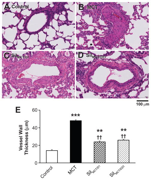
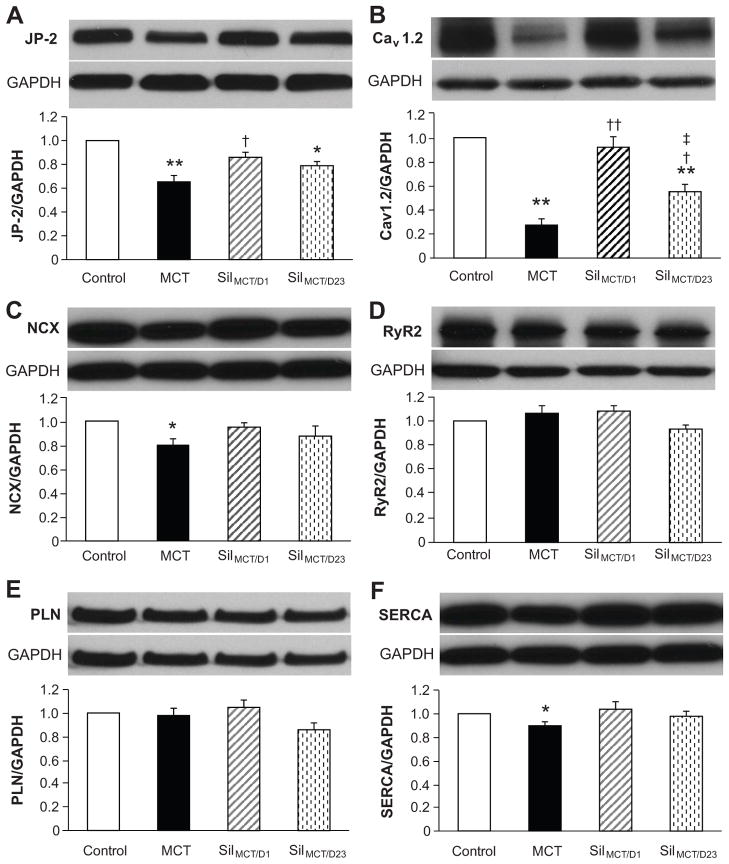
Right ventricular (RV) failure (RVF) is the main cause of death in patients with pulmonary artery hypertension (PAH). Sildenafil, a phosphodiesterase type 5 inhibitor, was approved recently for treatment of PAH patients. However, the mechanisms underlying RV contractile malfunction and the benefits of sildenafil on RV function are not well understood. We aimed to investigate the following: (1) the ultrastructural and excitation-contraction coupling alterations underlying PAH-induced RVF; (2) whether the ultrastructural changes are reversible; and (3) the mechanisms underlying the therapeutic benefits of sildenafil in PAH-RVF. We used a single injection of monocrotaline in Wistar rats to induce pulmonary vascular proliferation, which led to PAH and RVF. RV myocytes displayed severe transverse (T)-tubule loss and disorganization, as well as blunted and dys-synchronous sarcoplasmic reticulum Ca(2+) release. Sildenafil prevented and reversed the monocrotaline-induced PAH and LV filling impairment. Early intervention with sildenafil prevented RV hypertrophy and the development of RVF, T-tubule remodeling, and Ca(2+) handling dysfunction. Although late treatment with sildenafil did not reverse RV hypertrophy in animals with established RVF, RV systolic function was improved. Furthermore, late intervention partially reversed both the impairment of myocyte T-tubule integrity and Ca(2+) handling protein and sarcoplasmic reticulum Ca(2+) release function in monocrotaline-treated rats. In conclusion, PAH-induced increase in RV afterload causes severe T-tubule remodeling and Ca(2+) handling dysfunction in RV myocytes, leading to RV contractile failure. Sildenafil prevents and partially reverses ultrastructural, molecular, and functional remodeling of failing RV myocytes. Reversal of pathological T-tubule remodeling, although incomplete, is achievable without the regression of RV hypertrophy.


Conflict of interest statement

Disclosures

No conflict of interest to be disclosed.

Figures

Figure 1
Figure 1. Effects of MCT and sildenafil on vascular wall thickness in lung pulmonary arteries
A–D, Representative images of pulmonary vessels in the lungs of control (A), MCT (B), MCT rats treated with sildenafil beginning at day 1 (C) and MCT rats treated with sildenafil beginning at day 23 for two weeks (D). E, Vascular wall thickness in pulmonary vessels under 500 μm in length. **, p < 0.01; ***, p<0.001 vs. saline control; ††, p < 0.01 vs. MCT group. N=3, 3, 4, 4 lungs for each group, respectively.
Figure 2
Figure 2. Effects of MCT and sildenafil treatments on RV morphometric and RV systolic function
A–B, RV weight was measured after 24 hours of drying at 37°C (A) and normalized to LV+septum weight (B). N=5, 6, 7, 5 hearts per group, respectively. C–D, RV dimensions during systole (C) and diastole (D), and RV wall thickness during diastole (E). F, RV fractional shortening (FS, %), calculated as (RV dimension during diastole – RV dimension during systole) divided by RV dimension during diastole. N=11, 10, 9, 8 per group, respectively. MCT treated rats developed RVF with significant reduction in RV FS. Sildenafil early treatment prevented MCT induced RVF with maintained FS. Sildenafil late treatment improved RV systolic function, comparing to those of MCT alone. **, p < 0.001 vs. saline control; †, p<0.05; ††, p < 0.001 vs. MCT group; ‡, p<0.05 vs. SilMCT/D1 group.
Figure 3
Figure 3. MCT-induced PAH impairs LV filling
A, End diastolic volume; B, LV stroke volume; C, cardiac output. MCT induced PAH - RV afterload increase caused impairment of LV filling. Both early and late treatments with sildenafil normalized LV filling and output. D, LV ejection fraction (EF) was normal among all groups. ***, p<0.001 vs. saline control; †, p < 0.05; ††, p < 0.01 vs. MCT group. N=11, 10, 9, 8 per group, respectively.
Figure 4
Figure 4. Confocal imaging of Ca2+ transients in single isolated RV myocytes
A–D, representative Ca2+ transients from control, MCT, SilMCT/D1, and SilMCT/D23 groups, respectively. Shown underneath each panel of Ca2+ image is the spatially averaged Ca2+ profile. RV myocyte from MCT-treated rat (B) exhibited defects in the activation of Ca2+ transients (red arrows), prolonged plateau (green doublehead arrows) and slower decay phase (pink arrows). E–G, Summary data on parameters of Ca2+ transients, including amplitude (F/F0), time to peak (Tpeak) and decay rate (time to 50% decay, T50). *, p<0.05 vs control; **, p<0.01 vs control; †, p<0.05 vs MCT; ††, p<0.01 vs MCT. n=30, 54, 61, 52 cells (from 3–6 hearts), respectively.
Figure 5
Figure 5. In situ confocal imaging of RV myocyte T-tubule system from Langendorff perfused intact hearts
A–D, representative T-tubule images from control, MCT, SilMCT/D1, and SilMCT/D23 group, respectively. B, MCT-treated rats exhibited severe T-tubule loss and disorganization in RV myocytes. C, Early treatment of sildenafil (SilMCT/D1) prevented MCT-induced T-tubule remodeling. D, Late treatment of sildenafil (SilMCT/D23) partially reversed MCT induced T-tubule remodeling. E. Average data of TTpower (T-tubule power, an index of the strength of T-tubule regularity, for analysis, See reference Wei S, et al, 2010). TTpower of SilMCT/D1 group is not different from control. TTpower of SilMCT/D23 group is significantly higher than that of MCT rats, but lower than those of control and SilMCT/D1 groups. **, p<0.01 vs control; ††, p<0.01 vs MCT; ‡, p<0.05 vs. SilMCT/D1 group. N=7, 9, 7, 5 hearts per group, respectively.
Figure 6
Figure 6. Alterations in expression of Ca2+ handling proteins in RV myocytes in response to sildenafil treatment
RV tissue lysates were extracted for Western blotting assay. Representative blots and quantitation of JP-2 (A), Cav1.2 (B), NCX1 (C), RyR2 (D), PLN (E) and SERCA (F) levels. *, p<0.05 vs control; **, p<0.01 vs control; †, p<0.05 vs MCT; ††, p<0.01 vs MCT; ‡, p<0.05 vs. SilMCT/D1 group. N=4–5 hearts for each group.

Similar articles

Cited by

References

    1. Louch WE, Mork HK, Sexton J, Stromme TA, Laake P, Sjaastad I, Sejersted OM. T-tubule disorganization and reduced synchrony of Ca2+ release in murine cardiomyocytes following myocardial infarction. J Physiol. 2006;574:519–533. - PMC - PubMed
    1. Song LS, Sobie EA, McCulle S, Lederer WJ, Balke CW, Cheng H. Orphaned ryanodine receptors in the failing heart. Proc Natl Acad Sci USA. 2006;103:4305–4310. - PMC - PubMed
    1. Heinzel FR, Bito V, Biesmans L, Wu M, Detre E, von Wegner F, Claus P, Dymarkowski S, Maes F, Bogaert J, Rademakers F, D’Hooge J, Sipido K. Remodeling of t-tubules and reduced synchrony of Ca2+ release in myocytes from chronically ischemic myocardium. Circ Res. 2008;102:338–346. - PubMed
    1. Wei S, Guo A, Chen B, Kutschke W, Xie YP, Zimmerman K, Weiss RM, Anderson ME, Cheng H, Song LS. T-tubule remodeling during transition from hypertrophy to heart failure. Circ Res. 2010;107:520–531. - PMC - PubMed
    1. Balijepalli RC, Lokuta AJ, Maertz NA, Buck JM, Haworth RA, Valdivia HH, Kamp TJ. Depletion of t-tubules and specific subcellular changes in sarcolemmal proteins in tachycardia-induced heart failure. Cardiovasc Res. 2003;59:67–77. - PubMed

Publication types

MeSH terms