. 2011:2011:812135.
doi: 10.1155/2011/812135. Epub 2011 Dec 12.

The application of three-dimensional collagen-scaffolds seeded with myoblasts to repair skeletal muscle defects

Affiliations

The application of three-dimensional collagen-scaffolds seeded with myoblasts to repair skeletal muscle defects

Jianqun Ma et al. J Biomed Biotechnol. 2011.

Abstract

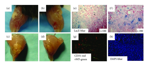
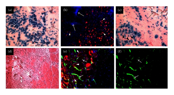
Three-dimensional (3D) engineered tissue constructs are a novel and promising approach to tissue repair and regeneration. 3D tissue constructs have the ability to restore form and function to damaged soft tissue unlike previous methods, such as plastic surgery, which are able to restore only form, leaving the function of the soft tissue often compromised. In this study, we seeded murine myoblasts (C2C12) into a collagen composite scaffold and cultured the scaffold in a roller bottle cell culture system in order to create a 3D tissue graft in vitro. The 3D graft created in vitro was then utilized to investigate muscle tissue repair in vivo. The 3D muscle grafts were implanted into defect sites created in the skeletal muscles in mice. We detected that the scaffolds degraded slowly over time, and muscle healing was improved which was shown by an increased quantity of innervated and vascularized regenerated muscle fibers. Our results suggest that the collagen composite scaffold seeded with myoblasts can create a 3D muscle graft in vitro that can be employed for defect muscle tissue repair in vivo.


Figures

Figure 1
Figure 1
Cell growth examined after 1 week of culturing in vitro. Cells were visualized throughout the scaffold with a higher concentration of cells located on the periphery of the scaffolds; however, extensive migration is seen within the scaffolds. H&E staining of the scaffolds ((a)–(c)), and LacZ and eosin staining ((d)–(f)) are shown.
Figure 2
Figure 2
Immunofluorescent staining of the scaffolds after culturing for 1, 2, and 3 weeks in vitro for evidence of vasculature ((a)–(c)). After one week of culturing (a), CD31 positive cells (red) are detected within the scaffold. After two weeks of culturing (b), colocalization of CD31 (red) and vWF (green) positive cells are visualized. After three weeks of culturing (c), more extensive colocalization of CD31 and vWF is seen. A BrdU assay was performed in vitro after 10 days of culturing ((d)–(f)). BrdU positive nuclei (red) were seen revealing that proliferation was occurring throughout the scaffold (e).
Figure 3
Figure 3
The immunocompatibility of the scaffolds was examined in normal mice and SCID mice in vivo. Twenty days after implantation in the normal mouse model, nonseeded scaffolds were rejected by the host tissue (a). Twenty days after implantation in the SCID mouse model, nonseeded scaffolds were not rejected by the host tissue, and few host cells migrated into the scaffold (b). Twenty days after implantation in the SCID mouse model, LacZ-labeled C2C12 myoblast seeded scaffolds were well received in the host tissue (c). Also, LacZ+ cells were able to migrate into the surrounding host tissue. An immunofluorescent staining of CD31 (red) and vWF (green) was performed on the implanted seeded scaffolds 20 days after implantation (d).
Figure 4
Figure 4
In the mdx/SCID mouse model, healthy healing was observed in the myoblast seeded scaffolds after 10 days (a) and 20 days (c). Nonseeded scaffolds implanted into mdx/SCID mice resulted in slower and less healthy healing at 10 days (b) and 20 days (d). LacZ staining of the implanted muscle graft region revealed extensive migration of the LacZ+ myoblasts into the surrounding host tissue (e) and (f). Immunofluorescent staining of CD31 (red) and vWF (green) showed vascularization developing in the implanted muscle graft region 20 days after implantation (g). A DAPI staining revealed that a variety of cell types were located homogenously throughout the implanted muscle graft area (h).
Figure 5
Figure 5
LacZ and eosin staining 1 month after implantation revealed that numerous LacZ+ myoblasts were able to survive within the implanted muscle graft region ((a) and (c)). Residues of nondegraded scaffold were also visualized within the implanted muscle graft area ((c), arrowheads). LacZ+ myoblasts were seen forming multinucleated myotubes within the implanted area ((a) arrows). Desmin positive myoblasts ((b) arrowheads) were detected within the vicinity of nondegraded portions of the scaffold ((b) arrows). An H&E staining revealed newly formed myofibers within the implantation site (d). Dystrophin-positive myofibers (red, arrowheads) were detected 1 month after implantation (e) as well as CD31-positive cells, ((e) and (f), green).
Figure 6
Figure 6
(a) Three months after implantation in vivo, large dystrophin-positive grafts were visualized ((a) B, E, H). CD31-positive cells were detected within the dystrophin-positive grafts indicative that vascularization and regeneration were occurring ((a) A–C). Neurofilament protein was visualized within the dystrophin-positive grafts revealing that innervations were forming along with the regenerating myofibers ((a) D–F). The majority of the dystrophin-positive grafts stained positively for fast myosin-heavy chain protein ((a) G–I). (b) Three months after implantation in vivo, all newly formed myofibers stained positive for dystrophin ((b) A dystrophin-red). A small quantity of the dystrophin-positive myofibers stained positive for slow myosin-heavy chain protein ((b) B slow MyHC-gray). A larger quantity of the dystrophin-positive myofibers stained positive for fast myosin-heavy chain protein ((b) C fast MyHC-green). (b) D displays a merged image revealing the distribution of the two types of myosin-heavy chain proteins.
Figure 7
Figure 7
Three months after implantation, evaluation of the regenerative capacity of the seeded scaffolds. A hematoxylin and eosin staining revealed a large area of fibrosis in the muscle graft ((a) and (b), sk). However, within the muscle graft despite the area of fibrosis, we detected regenerating myofibers ((b), arrows), and large quantities of dystrophin-positive myofibers ((c), arrows). Within the areas of regeneration, revascularization was detected through the visualization of CD31-positive cells ((d) green/arrows).

Similar articles

Cited by

References

    1. Dragas M, Davidovic L, Kostic D, et al. Upper extremity arterial injuries: factors influencing treatment outcome. Injury. 2009;40(8):815–819. - PubMed
    1. Attinger CE, Ducic I, Cooper P, Zelen CM. The role of intrinsic muscle flaps of the foot for bone coverage in foot and ankle defects in diabetic and nondiabetic patients. Plastic and Reconstructive Surgery. 2002;110(4):1047–1054. - PubMed
    1. Singh AK, Gudehithlu KP, Patri S, et al. Impaired integration of endothelial progenitor cells in capillaries of diabetic wounds is reversible with vascular endothelial growth factor infusion. Translational Research. 2007;149(5):282–291. - PubMed
    1. Al-Qattan MM. Severe, traumatic soft-tissue loss in the antecubital fossa and proximal forearm associated with radial and/or median nerve palsy: nerve recovery after coverage with a pedicled latissimus dorsi muscle flap. Annals of Plastic Surgery. 2001;46(2):125–129. - PubMed
    1. Takeuchi A, Tsuchiya H, Shirai T, Hayashi K, Nishida H, Tomita K. Occlusive dressing for large soft tissue defects following soft tissue tumor excision. Journal of Orthopaedic Science. 2009;14(4):385–390. - PubMed

LinkOut - more resources