

Ixora parviflora Protects against UVB-Induced Photoaging by Inhibiting the Expression of MMPs, MAP Kinases, and COX-2 and by Promoting Type I Procollagen Synthesis
- PMID: 22203872
- PMCID: PMC3235733
- DOI: 10.1155/2012/417346
Ixora parviflora Protects against UVB-Induced Photoaging by Inhibiting the Expression of MMPs, MAP Kinases, and COX-2 and by Promoting Type I Procollagen Synthesis
Abstract
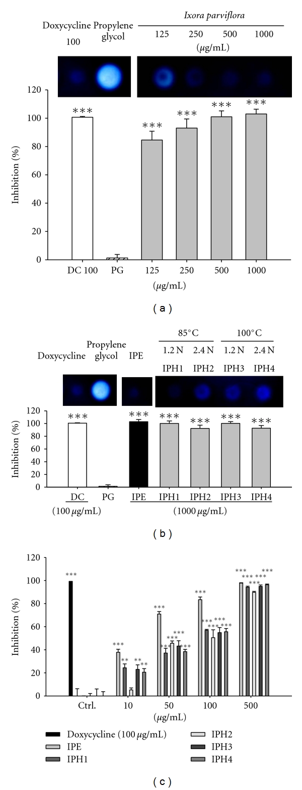
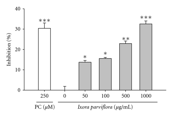
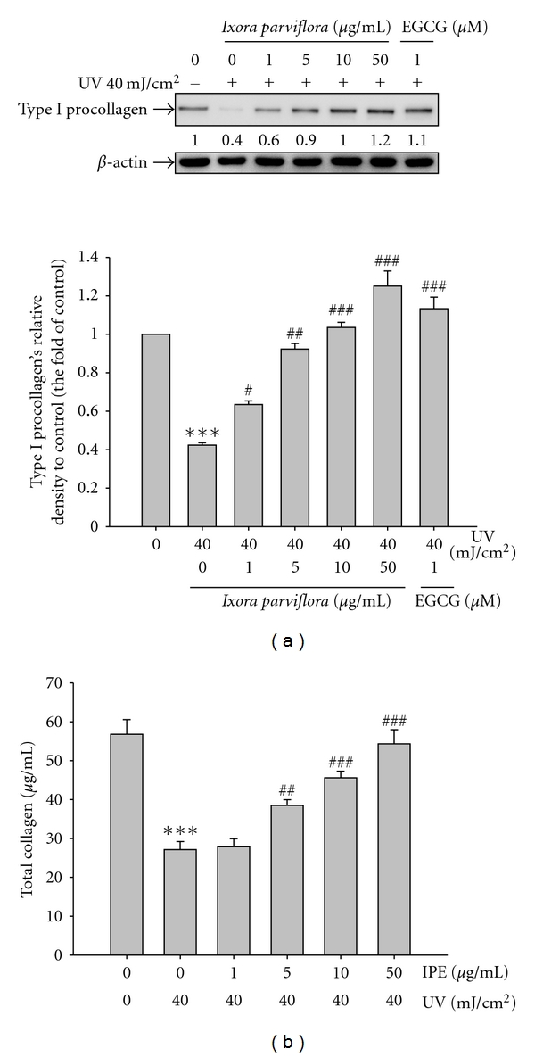
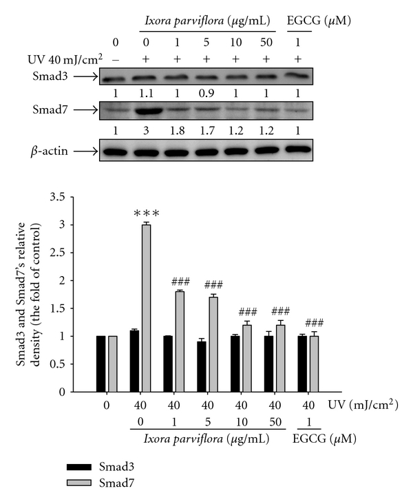
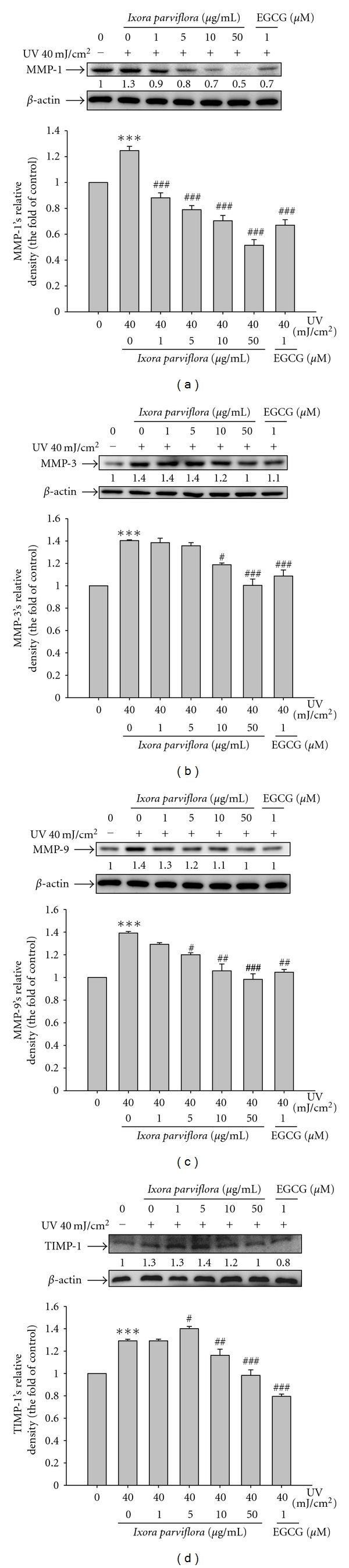
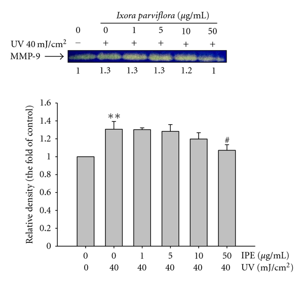
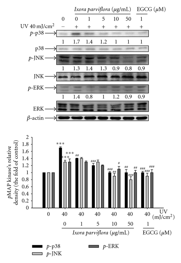
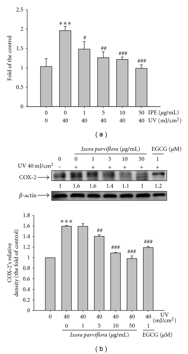
Ixora parviflora with high polyphenol content exhibited antioxidant activity and reducing UVB-induced intracellular reactive oxygen species production. In this study, results of the photoaging screening experiments revealed that IPE at 1000 μg/mL reduced the activity of bacterial collagenase by 92.7 ± 4.2% and reduced the activity of elastase by 32.6 ± 1.4%. Therefore, we investigated the mechanisms by which IPE exerts its anti-photoaging activity. IPE at 1 μg/mL led to an increase in type I procollagen expression and increased total collagen synthesis in fibroblasts at 5 μg/mL. We found that IPE inhibited MMP-1, MMP-3, and MMP-9 expression at doses of 1, 5, and 10 μg/mL, respectively, in fibroblasts exposed to UV irradiation (40 mJ/cm(2)). Gelatin zymography assay showed that IPE at 50 μg/mL inhibited MMP-9 secretion/activity in cultured fibroblasts after UVB exposure. In addition, IPE inhibited the phosphorylation of p38, ERK, and JNK induced by UVB. Furthermore, IPE inhibited the UVB-induced expression of Smad7. In addition, IPE at 1 μg/mL inhibited NO production and COX-2 expression in UV-exposed fibroblasts. These findings show that IPE exhibits anti-inflammatory and anti-photoaging activities, indicating that IPE could be a potential anti-aging agent.
Figures
Similar articles
-
Pinus densiflora extract protects human skin fibroblasts against UVB-induced photoaging by inhibiting the expression of MMPs and increasing type I procollagen expression.Toxicol Rep. 2014 Aug 29;1:658-666. doi: 10.1016/j.toxrep.2014.08.010. eCollection 2014. Toxicol Rep. 2014. PMID: 28962279 Free PMC article.
-
Michelia alba extract attenuates UVB-induced expression of matrix metalloproteinases via MAP kinase pathway in human dermal fibroblasts.Food Chem Toxicol. 2012 Dec;50(12):4260-9. doi: 10.1016/j.fct.2012.08.018. Epub 2012 Aug 17. Food Chem Toxicol. 2012. PMID: 22922035
-
Antioxidant activity of Ixora parviflora in a cell/cell-free system and in UV-exposed human fibroblasts.Molecules. 2011 Jul 6;16(7):5735-52. doi: 10.3390/molecules16075735. Molecules. 2011. PMID: 21734630 Free PMC article.
-
Anti-Photoaging Effect of Phaseolus angularis L. Extract on UVB-Exposed HaCaT Keratinocytes and Possibilities as Cosmetic Materials.Molecules. 2023 Feb 1;28(3):1407. doi: 10.3390/molecules28031407. Molecules. 2023. PMID: 36771069 Free PMC article.
-
Oleracone C from Portulaca oleracea attenuates UVB-induced changes in matrix metalloproteinase and type I procollagen production via MAPK and TGF-β/Smad pathways in human keratinocytes.Int J Cosmet Sci. 2023 Apr;45(2):166-176. doi: 10.1111/ics.12828. Epub 2023 Jan 12. Int J Cosmet Sci. 2023. PMID: 36415152
Cited by
-
Taxifolin as a Promising Ingredient of Cosmetics for Adult Skin.Antioxidants (Basel). 2021 Oct 15;10(10):1625. doi: 10.3390/antiox10101625. Antioxidants (Basel). 2021. PMID: 34679758 Free PMC article.
-
Dieckol Isolated from Eisenia bicyclis Ameliorates Wrinkling and Improves Skin Hydration via MAPK/AP-1 and TGF-β/Smad Signaling Pathways in UVB-Irradiated Hairless Mice.Mar Drugs. 2022 Dec 14;20(12):779. doi: 10.3390/md20120779. Mar Drugs. 2022. PMID: 36547926 Free PMC article.
-
Skin ageing: natural weapons and strategies.Evid Based Complement Alternat Med. 2013;2013:827248. doi: 10.1155/2013/827248. Epub 2013 Jan 29. Evid Based Complement Alternat Med. 2013. PMID: 23431351 Free PMC article.
-
Hydrangea serrata (Thunb.) Ser. Extract Attenuate UVB-Induced Photoaging through MAPK/AP-1 Inactivation in Human Skin Fibroblasts and Hairless Mice.Nutrients. 2019 Mar 1;11(3):533. doi: 10.3390/nu11030533. Nutrients. 2019. PMID: 30823635 Free PMC article.
-
Protective Activity and Underlying Mechanism of Ginseng Seeds against UVB-Induced Damage in Human Fibroblasts.Antioxidants (Basel). 2021 Mar 8;10(3):403. doi: 10.3390/antiox10030403. Antioxidants (Basel). 2021. PMID: 33800272 Free PMC article.
References
-
- Chung JH. Photoaging in Asians. Photodermatology, Photoimmunology & Photomedicine. 2003;19:109–121. - PubMed
-
- Ley RD, Peak MJ, Lyon LL. Induction of pyrimidine dimers in epidermal DNA of hairless mice by UVB: an action spectrum. Journal of Investigative Dermatology. 1983;80(3):188–191. - PubMed
-
- Pfeifer GP, You YH, Besaratinia A. Mutations induced by ultraviolet light. Mutation Research. 2005;571(1-2):19–31. - PubMed
-
- Marrot L, Meunier JR. Skin DNA photodamage and its biological consequences. Journal of the American Academy of Dermatology. 2008;58(5):S139–S148. - PubMed
-
- Gelse K, Pöschl E, Aigner T. Collagens—structure, function, and biosynthesis. Advanced Drug Delivery Reviews. 2003;55(12):1531–1546. - PubMed
LinkOut - more resources
Full Text Sources
Medical
Research Materials
Miscellaneous
