. 2012 Jan 10;109(2):616-20.
doi: 10.1073/pnas.1119751109. Epub 2011 Dec 27.

AMP-activated protein kinase is physiologically regulated by inositol polyphosphate multikinase

Affiliations

AMP-activated protein kinase is physiologically regulated by inositol polyphosphate multikinase

Sookhee Bang et al. Proc Natl Acad Sci U S A. .

Abstract

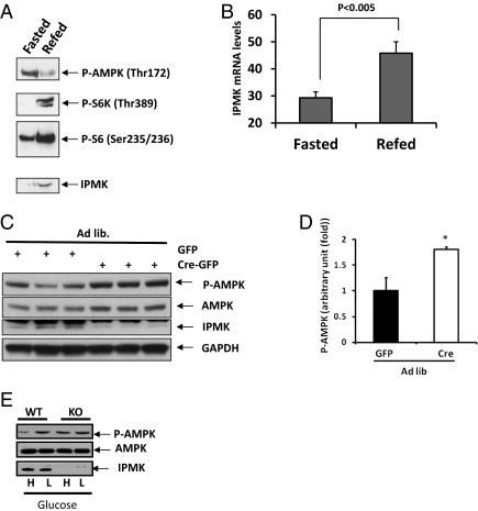
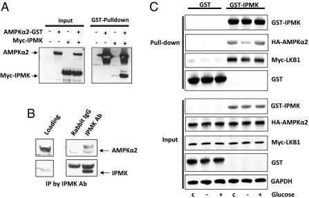
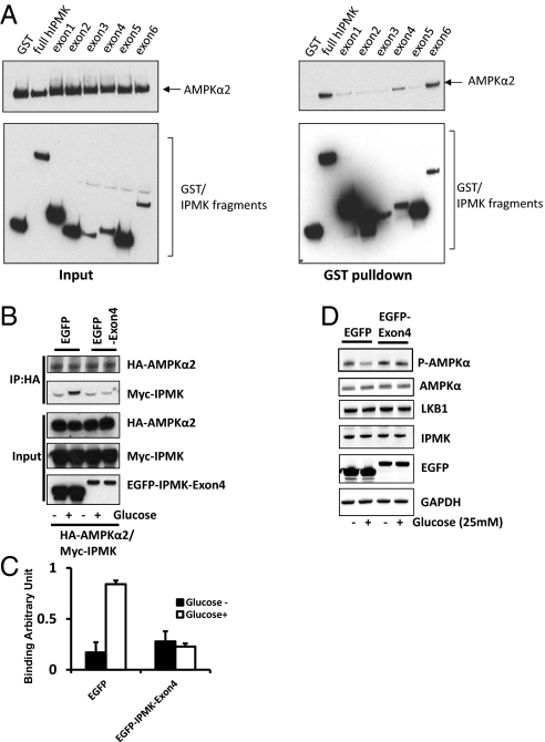
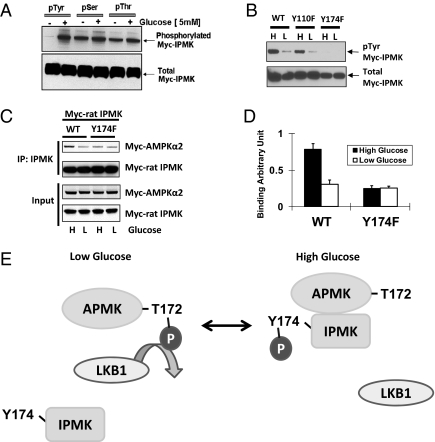
The AMP-activated kinase (AMPK) senses the energy status of cells and regulates fuel availability, whereas hypothalamic AMPK regulates food intake. We report that inositol polyphosphate multikinase (IPMK) regulates glucose signaling to AMPK in a pathway whereby glucose activates phosphorylation of IPMK at tyrosine 174 enabling the enzyme to bind to AMPK and regulate its activation. Thus, refeeding fasted mice rapidly and markedly stimulates transcriptional enhancement of IPMK expression while down-regulating AMPK. Also, AMPK is up-regulated in mice with genetic depletion of hypothalamic IPMK. IPMK physiologically binds AMPK, with binding enhanced by glucose treatment. Regulation by glucose of phospho-AMPK in hypothalamic cell lines is prevented by blocking AMPK-IPMK binding. These findings imply that IPMK inhibitors will be beneficial in treating obesity and diabetes.


Conflict of interest statement

The authors declare no conflict of interest.

Figures

Fig. 1.
Fig. 1.
IPMK physiologically regulates AMPK activation in response to nutrients. (A and B) Effects of refeeding on IPMK gene expression in the hypothalamus. Mice fasted for 24 h were refed for 2 h, the whole hypothalami were isolated, and RNA or protein extracts were prepared as described in Materials and Methods. (A) Westerns blots are shown for P-AMPKα, P-S6K, P-S6, and IPMK. (B) Quantification of IPMK mRNA levels in fasted and refed mice (n = 5 per group). (C and D) Loss of hypothalamic IPMK leads to elevated P-AMPKα in ad libitum mice. IPMKlox/lox mice were injected with adenovirus expressing either GFP or Cre recombinase. Mice were fed ad libitum and killed. (C) Westerns blots are shown of P-AMPKα, AMPK, IPMK, and GAPDH. (D) Relative quantifications of P-AMPKα expression levels are shown in GFP-infected (black bar) or GFP-Cre-infected (open bar) mice. Values are corrected for corresponding total AMPK antibody (*Student t test; P < 0.005). (E) IPMK flox/flox (WT) and IPMK−/− (KO) MEFs were incubated with DMEM media containing either high glucose (4.5 g/L) or low glucose (1.5 g/L).
Fig. 2.
Fig. 2.
IPMK regulates AMPK activation through interaction of IPMK-AMPK in response to glucose availability. (A) IPMK interacts with AMPKα2. GST-AMPKα2 was cotransfected in HEK293T cells with myc-IPMK, followed by GST pull-down assay. The precipitated proteins and the input proteins were detected by immunoblotting with antibodies to GST or myc. (B) Hypothalamic lysate (500 μg) was used for immunoprecipitation (IP) against Rabbit IgG antibody or IPMK antibody to determine the physiological binding of AMPKα2. (C) HA-AMPKα2 and Myc-LKB1 were cotransfected in GT1-7 cells with GST or GST-IPMK as indicated. Cells were deprived of glucose for 3 h and stimulated with glucose for 30 min. GST pull-down assay was performed to determine IPMK and AMPKα2 interaction.
Fig. 3.
Fig. 3.
Physical interaction between IPMK and AMPK is required for IPMK-mediated modulation of AMPK. (A) Mapping of binding region of IPMK responsible for AMPK interaction. GST, GST-IPMK or GST–IPMK exon fragments (exon 1: 1–62, exon 2: 63–92, exon 3: 93–124, exon 4: 125–182, exon 5: 183–208 and exon 6: 209–416) were pull-downed from HEK293T cells cotransfected with AMPKα2. Coimmunoprecipitation of AMPKα2 was determined by Western blots. (BD) Dominant-negative IPMK exon 4 disrupts the IPMK-AMPK interaction. (B) HA-AMPKα2, Myc-IPMK, and EGFP or EGFP-IPMK exon 4 were cotransfected into GT1-7 cells as indicated. Cells were deprived of glucose for 3 h and stimulated with glucose for 30 min before lysis. HA-AMPKα2 was immunoprecipitated and coimmunoprecipitates of Myc-IPMK were determined by Western blot (C) Relative quantifications of bound AMPKα2 and IPMK levels are shown. Values are expressed as means ± SD of three determinations (*Student t test; P < 0.005). (D) EGFP or EGFP-exon 4 was transfected into HEK293 cells as indicated. Cells were deprived of glucose for 3 h and stimulated with or without glucose for 30 min before lysis. Proteins were extracted and analyzed by Western blotting.
Fig. 4.
Fig. 4.
IPMK phosphorylation is required for AMPK interaction on the high glucose. (A) High glucose induces IPMK phosphorylation. GT1-7 cells were transfected with Myc rat-IPMK. Cells were starved of glucose for 3 h and resupplied with glucose (5 mM) for 30 min. Phosphorylated protein was immunoprecipitated with phospho-tyrosine, phospho-serine, and phospho-threonine antibodies, respectively. Phosphorylated (top) and total (bottom) IPMK was determined by Western blot. (B) Comparisons of tyrosine phosphorylated IPMK in cells overexpressed WT and tyrosine mutants of rat IPMK. WT or two tyrosine mutants (Y110F and Y174F) were transfected into GT1-7 cells and incubated with DMEM containing high glucose (H) or low glucose (L) for 24 h. Immunoprecipitation was done with phospho-tyrosine antibody to the indicated proteins from lysates and Western blots of tyrosine-phosphorylated IPMK. (C) Myc-AMPKα2 and Myc-IPMK were cotransfected into GT1-7 cells as indicated. Cells were incubated with DMEM containing high glucose (H) or low glucose (L) for 24 h. IPMK was immunoprecipitated, and Western blots were performed. (D) Relative quantifications of bound AMPKα2 and IPMK levels are shown (*Student t test, P < 0.005). (E) Schematic representation of a model explaining how IPMK interacts with AMPK in response to glucose signals. In low glucose, tyrosine phosphorylation of IPMK is decreased, and, subsequently, AMPK can interact with and be phosphorylated by LKB1. In high glucose, increased phosphorylation of Y174 in IPMK interferes with the action of LKB1 to decrease phosphorylation of AMPK. Alternatively, IPMK may promote dephosphorylation of AMPK.

References

    1. Carling D. The AMP-activated protein kinase cascade—a unifying system for energy control. Trends Biochem Sci. 2004;29:18–24. - PubMed
    1. Hardie DG, Hawley SA, Scott JW. AMP-activated protein kinase—development of the energy sensor concept. J Physiol. 2006;574:7–15. - PMC - PubMed
    1. Mihaylova MM, Shaw RJ. The AMPK signalling pathway coordinates cell growth, autophagy and metabolism. Nat Cell Biol. 2011;13:1016–1023. - PMC - PubMed
    1. Hardie DG, Pan DA. Regulation of fatty acid synthesis and oxidation by the AMP-activated protein kinase. Biochem Soc Trans. 2002;30:1064–1070. - PubMed
    1. Minokoshi Y, et al. AMP-kinase regulates food intake by responding to hormonal and nutrient signals in the hypothalamus. Nature. 2004;428:569–574. - PubMed

Publication types

MeSH terms

Substances