. 2012 May 9;18(1):359-69.
doi: 10.2119/molmed.2011.00203.

Poly(ADP-ribosyl)ation of high mobility group box 1 (HMGB1) protein enhances inhibition of efferocytosis

Affiliations

Poly(ADP-ribosyl)ation of high mobility group box 1 (HMGB1) protein enhances inhibition of efferocytosis

Kasey Davis et al. Mol Med. .

Abstract

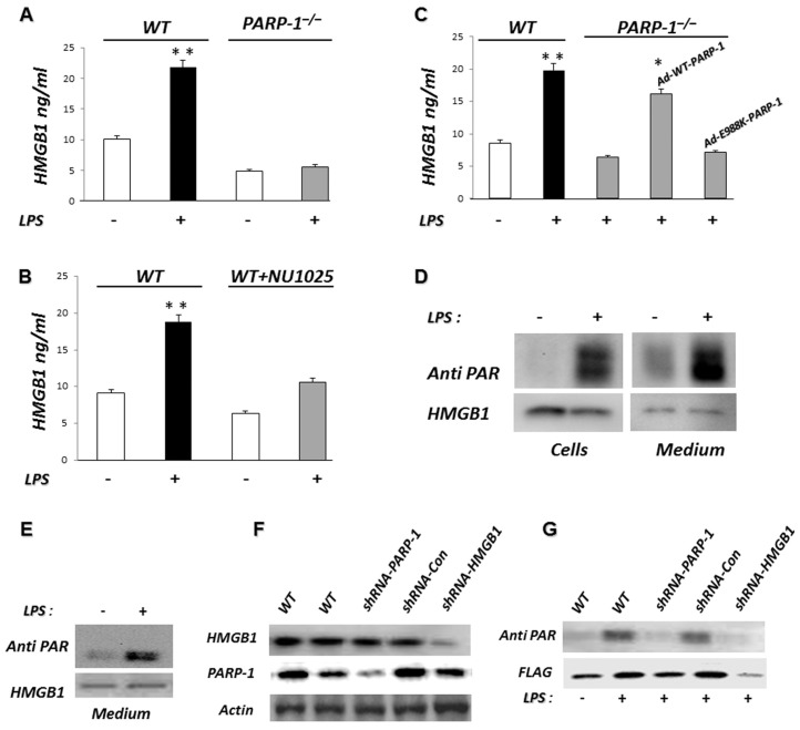
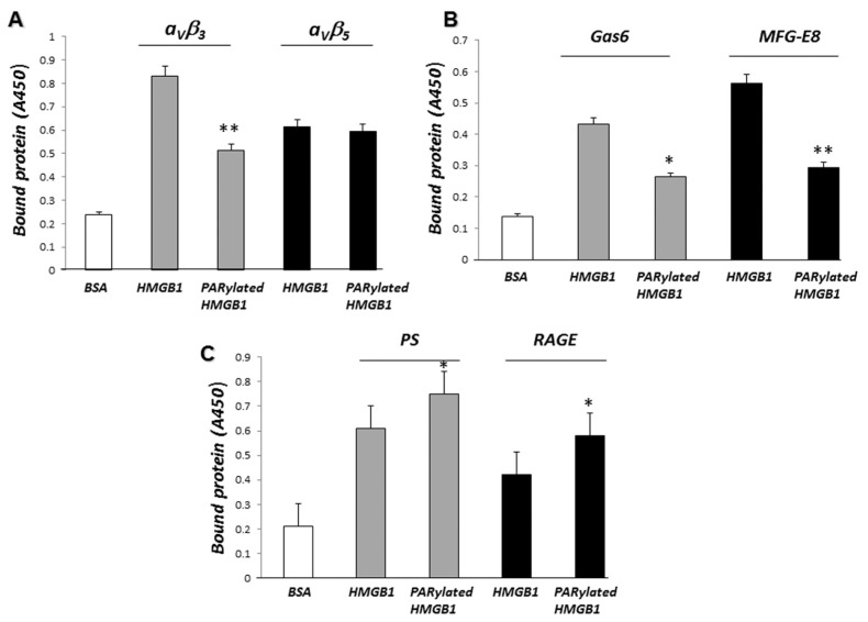
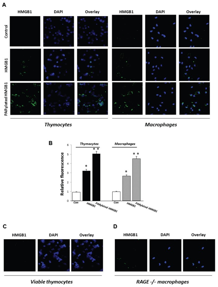
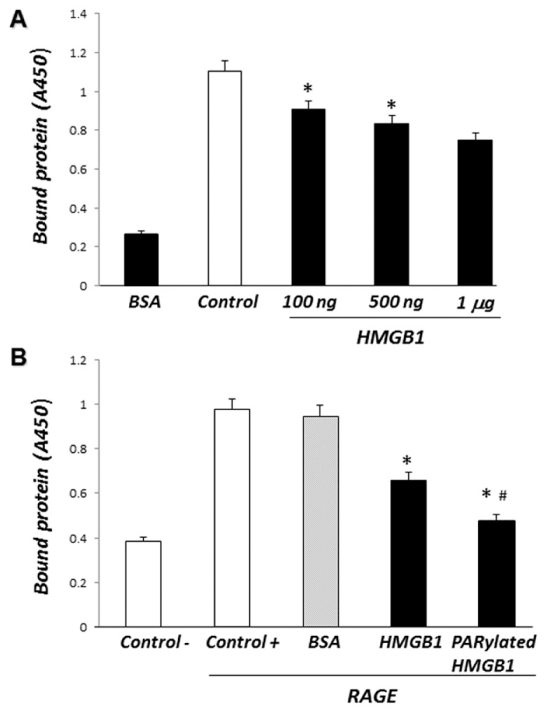
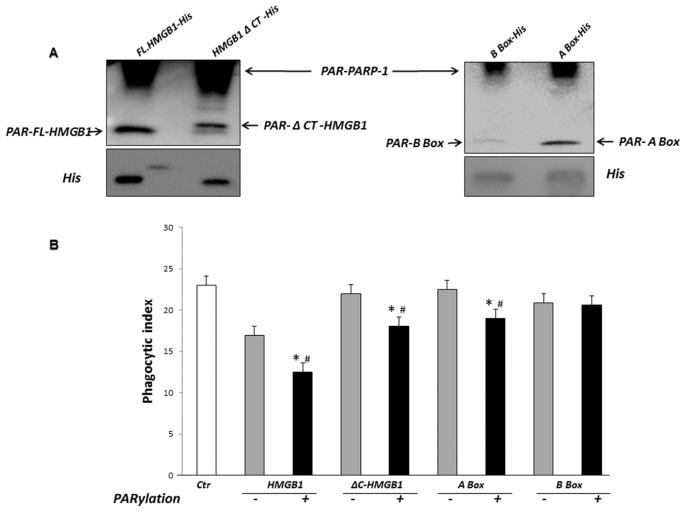
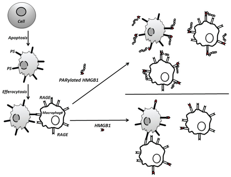
Phagocytosis of apoptotic cells by macrophages, known as efferocytosis, is a critical process in the resolution of inflammation. High mobility group box 1 (HMGB1) protein was first described as a nuclear nonhistone DNA-binding protein, but is now known to be secreted by activated cells during inflammatory processes, where it participates in diminishing efferocytosis. Although HMGB1 is known to undergo modification when secreted, the effect of such modifications on the inhibitory actions of HMGB1 during efferocytosis have not been reported. In the present studies, we found that HMGB1 secreted by Toll-like receptor 4 (TLR4) stimulated cells is highly poly(ADP-ribosyl)ated (PARylated). Gene deletion of poly(ADP)-ribose polymerase (PARP)-1 or pharmacological inhibition of PARP-1 decreased the release of HMGB1 from the nucleus to the extracellular milieu after TLR4 engagement. Preincubation of macrophages or apoptotic cells with HMGB1 diminished efferocytosis through mechanisms involving binding of HMGB1 to phosphatidylserine on apoptotic cells and to the receptor for advanced glycation end products (RAGE) on macrophages. Preincubation of either macrophages or apoptotic cells with PARylated HMGB1 inhibited efferocytosis to a greater degree than exposure to unmodified HMGB1, and PARylated HMGB1 demonstrated higher affinity for phosphatidylserine and RAGE than unmodified HMGB1. PARylated HMGB1 had a greater inhibitory effect on Ras-related C3 botulinum toxin substrate 1 (Rac-1) activation in macrophages during the uptake of apoptotic cells than unmodified HMGB1. The present results, showing that PARylation of HMGB1 enhances its ability to inhibit efferocytosis, provide a novel mechanism by which PARP-1 may promote inflammation.


Figures

Figure 1
Figure 1
Extracellular HMGB1 is highly PARylated in cells stimulated with LPS. (A) PARP-1 gene deletion inhibits LPS-induced HMGB1 release. WT or PARP-1−/− MEFs were treated with 100 ng/mL LPS for 24 h at 37°C and then extracellular HMGB1 was secreted into the culture supernatant quantified by ELISA. **P < 0.01 compared with nontreated cells. Assay was done in triplicate. (B) Inhibition of PARP-1 reduces LPS-induced HMGB1 release. WT MEF cells were treated with LPS in the presence or absence of the noncompetitive PARP-1 inhibitor NU1025 for 24 h, after which extracellular HMGB1 was quantified by ELISA. **P < 0.01 compared with nontreated cells. (C) The decrease in HMGB1 release by LPS-stimulated PARP-1−/− MEFs is reversed by reconstitution of expression of WT PARP-1 but not by enzymatically inactive PARP-1 (E988K). PARP-1−/−MEFs were infected with an adenoviral vector expressing EYFP-WT PARP-1 (Ad-WT-PARP-1) or EYFP-E988K PARP-1 (Ad-E988K-PARP-1) or left uninfected. Forty-eight hours later, the cells were cultured for 24 h with 100 ng/mL LPS or left untreated (Con). Extracellular HMGB1 was quantified by ELISA. *P < 0.05 compared with uninfected PARP-1−/− MEFs. **P < 0.01 compared with untreated WT MEFs. (D) HMGB1 secreted after TLR4 stimulation is highly PARylated. J447 mouse cells expressing Flag-HMGB1 were treated with 100 ng/mL LPS for 24 h or left untreated (Con). The culture supernatants and protein extracts prepared from the cells were then subjected to pull-down with anti-Flag agarose beads. The resulting precipitates were subjected to immunoblot analysis with antibodies to PAR and HMGB1. A representative gel is shown. Two additional independent experiments provided similar results. (E) Flag-HMGB1 pull-down under nondenaturing conditions and after PARP-1 and HMGB1 knockdown. J447 mouse cells expressing Flag-HMGB1 were treated with 100 ng/mL LPS for 24 h or left untreated (Con). The protein extracts were then subjected to pull-down with anti-Flag agarose beads under nondenaturing conditions. The resulting precipitates were subjected to immunoblot analysis with antibodies to PAR or HMGB1. (F) Flag-HMGB1 pull-down under denaturing conditions after PARP-1 and HMGB1 knockdown. The same experiment was conducted with J447-mouse cells expressing Flag-HMGB1 after PARP-1 or HMGB1 knockdown. The cells were transduced with lentiviruses encoding control shRNA (shRNA-Con), shRNA targeting HMGB1 (shRNA-HMGB1) or shRNA targeting PARP-1 (shRNA-PARP-1); expression of PARP-1, HMGB1 and actin was assessed by immunoblot analysis and compared with that in uninfected cells. (G) These infected or uninfected cells were treated with 100 ng/mL LPS for 24 h or left untreated. The protein extracts were then subjected to pull-down with anti-Flag agarose beads. The resulting precipitates were subjected to immunoblot analysis with antibodies to PAR or Flag. Results from representative experiments are shown. A second set of independent experiments provided similar findings.
Figure 2
Figure 2
PARylated HMGB1 inhibits phagocytosis of apoptotic cells by macrophages more effectively than non-PARylated HMGB1. (A) Phagocytosis assays were performed after preincubation of apoptotic thymocytes for 2 h with 1 μg/mL recombinant HMGB1 or PARylated HMGB1. *P < 0.05 and **P < 0.01 compared with control BSA-treated thymocytes. #P < 0.05 compared with HMGB1-treated thymocytes. (B) After preincubation of thymocytes for 2 h with PARylated or unmodified HMGB1, phagocytosis assays were performed for 5 and 30 min. The macrophages were then washed three times, and cell lysates were prepared to determine Rac-1 activation. As a negative control, macrophages were preincubated with media in the absence of thymocytes. White bars represent unmodified HMGB1, and gray bars represent PARylated HMGB1; *P < 0.05 versus PARylated HMGB1. (C) Phagocytosis assays were performed after preincubation of macrophages for 2 h with 1 μg/mL recombinant HMGB1 or PARylated HMGB1. *P < 0.05 compared with control BSA-treated macrophages. #P < 0.05 compared with HMGB1-treated macrophages. (D.1) Phagocytosis assays were performed after preincubation of macrophages for 2 h with 1 μg/mL PARylated HMGB1 or free PAR and compared with control BSA-treated macrophages. (D.2) PARP-1 protein was incubated in a poly(ADP-ribosyl)ation reaction containing NAD and activated DNA for 30 min. PARG was then added to the reaction for 30 min. Separation of free PAR from PARP-1 was performed using Amicon Ultra 0.5 filters. A portion (10%) of the reaction mixture was used for immunoblot analysis with antibodies to PAR. A representative gel is shown. A second independent experiment provided similar results. All phagocytosis experiments were performed at least three independent times with similar results. (E) Apoptotic thymocytes (107) preincubated for 2 h with 2 μg HMGB1, PARylated HMGB1 or BSA were administered intratracheally in 50 μL PBS into anesthesized mice. BAL fluid was collected 90 min later. The samples were resuspended in PBS with 1% BSA and stained with FITC-CD11b Ab (macrophage marker) and APC-CD 90.2 Ab (thymocyte marker). Flow cytometry was performed, and the phagocytosis index was calculated as the ratio of FITC+PKH26+APC cells to all cells gated. n = 3 mice in each group. *P < 0.05 compared with control. #P < 0.05 compared with HMGB1-treated thymocytes. A second independent experiment provided similar results.
Figure 3
Figure 3
PARylated HMGB1 binds to PS and RAGE with higher affinity than non-PARylated HMGB1. (A) After being precoated with the integrins αVβ3 or αVβ5, 96-well plates were incubated with 1 μg/mL HMGB1 or PARylated HMGB1 for 2 h. The plates were then washed and bound HMGB1 was quantified by ELISA, as described in Materials and Methods. **P < 0.01 compared with HMGB1. (B) Gas6 and MFG-E8–precoated 96-well plates were incubated with 1 μg/mL HMGB1 or PARylated HMGB1 for 2 h. The plates were then washed and bound HMGB1 was quantified by ELISA. *P < 0.05 and **P < 0.01 compared with HMGB1. (C) Recombinant RAGE dissolved in PBS or PS dissolved in methanol was incubated in 96-well plates. The plates were then incubated with 1 μg/mL HMGB1 or PARylated HMGB1 for 2 h. Bound HMGB1 was quantified by ELISA. *P < 0.05 compared with HMGB1. Assays were done in triplicate.
Figure 4
Figure 4
PARylated HMGB1 binds with increased affinity to PS on apoptotic cells and to RAGE on macrophages compared with unmodified HMGB1. (A) Apoptotic thymocytes or WT macrophages were incubated with 1 μg/mL HMGB1 or PARylated HMGB1 for 2 h at 37°C or left untreated (Control), and then HMGB1 binding was examined by immunofluorescence. Representative images are shown. Two additional independent experiments provided similar results. (B) Quantification of HMGB1 binding to apoptotic cells or macrophages using ImageJ software (n = 10 cells from three independent experiments). Means ± SEM are shown. *P < 0.05 and **P < 0.01 compared with control. (C, D) PARylated HMGB1 does not bind to viable thymocytes and binds with diminished affinity to RAGE−/− macrophages. Viable thymocytes (C) or RAGE−/− macrophages (D) were incubated with 1 μg/mL PARylated HMGB1 for 2 h at 37°C, and then HMGB1 binding was examined by immunofluorescence. Representative images are shown. Two additional independent experiments provided similar results.
Figure 5
Figure 5
PARylated HMGB1 inhibits the binding of RAGE to PS more effectively than non-PARylated HMGB1. (A) PS-precoated 96-well plates were incubated with increasing concentrations of recombinant HMGB1 (100 ng/mL, 500 ng/mL or 1 μg/mL) for 2 h at room temperature. Recombinant mouse chimeric RAGE was then added to the wells for 2 h. The plates were washed and bound RAGE was quantified by ELISA. All assays were performed in triplicate. *P < 0.05 compared with control. (B) PS-precoated 96-well plates were incubated with recombinant HMGB1, PARylated HMGB1 or BSA (1 μg/mL) for 2 h at room temperature. Recombinant mouse chimeric RAGE was then added to the wells for 2 h. The plates were washed and bound RAGE was quantified by ELISA. All assays were done in triplicate. *P < 0.05 compared with control. #P < 0.05 compared with HMGB1.
Figure 6
Figure 6
Poly(ADP-ribosyl)ation by PARP-1 of HMGB1 lacking the C-terminal tail and of HMGB1 A-Box, but not HMGB1 B-Box, potentiates the inhibitory effects of HMGB1 on efferocytosis. (A) PARylation of full-length HMGB1 and HMGB1 domains by PARP-1. Full-length HMGB1-His, ΔC-HMGB1-His, A Box-His or B Box-His (500 ng/mL) were incubated with recombinant PARP-1 in a poly(ADP-ribosyl)ation reaction containing NAD and activated DNA for 30 min. The reactions were terminated with sample buffer and subjected to immunoblot analysis with antibodies to PAR or His. Representative gels are shown. Two additional independent experiments provided similar results. (B) Phagocytosis assays were performed after preincubation of apoptotic thymocytes with 1 μg/mL unmodified or PARylated HMGB1-His, ΔC-HMGB1-His, A Box-His or B Box-His for 2 h. The thymocytes were then washed before being added to macrophages. *P < 0.05 compared with control. #P < 0.05 compared with thymocytes treated with non-PARylated HMGB1 or HMGB1 domains.
Figure 7
Figure 7
Proposed mechanism by which PARylation enhances HMGB1-associated inhibition of efferocytosis. PARylation increases the ability of HMGB1 to inhibit efferocytosis by binding more efficiently to PS on apoptotic cells and to RAGE on macrophages, thereby diminishing association of the apoptotic cell with the macrophage by interfering with binding between RAGE and PS.

Similar articles

Cited by

References

    1. Haslett C. Granulocyte apoptosis and its role in the resolution and control of lung inflammation. Am J Respir Crit Care Med. 1999;160:S5–11. - PubMed
    1. Voll RE, et al. Immunosuppressive effects of apoptotic cells. Nature. 1997;390:350–1. - PubMed
    1. Fadok VA, et al. Macrophages that have ingested apoptotic cells in vitro inhibit proinflammatory cytokine production through autocrine/paracrine mechanisms involving TGF-beta, PGE2, and PAF. J Clin Invest. 1998;101:890–8. - PMC - PubMed
    1. Ulloa L, Batliwalla FM, Andersson U, Gregersen PK, Tracey KJ. High mobility group box chromosomal protein 1 as a nuclear protein, cytokine, and potential therapeutic target in arthritis. Arthritis Rheum. 2003;48:876–81. - PubMed
    1. Czura CJ, Yang H, Amella CA, Tracey KJ. HMGB1 in the immunology of sepsis (not septic shock) and arthritis. Adv Immunol. 2004;84:181–200. - PubMed

Publication types

MeSH terms