. 2011 Dec 28:9:32.
doi: 10.1186/1478-811X-9-32.

The signaling pathway of Campylobacter jejuni-induced Cdc42 activation: Role of fibronectin, integrin beta1, tyrosine kinases and guanine exchange factor Vav2

Affiliations

The signaling pathway of Campylobacter jejuni-induced Cdc42 activation: Role of fibronectin, integrin beta1, tyrosine kinases and guanine exchange factor Vav2

Malgorzata Krause-Gruszczynska et al. Cell Commun Signal. .

Abstract

Background: Host cell invasion by the foodborne pathogen Campylobacter jejuni is considered as one of the primary reasons of gut tissue damage, however, mechanisms and key factors involved in this process are widely unclear. It was reported that small Rho GTPases, including Cdc42, are activated and play a role during invasion, but the involved signaling cascades remained unknown. Here we utilised knockout cell lines derived from fibronectin-/-, integrin-beta1-/-, focal adhesion kinase (FAK)-/- and Src/Yes/Fyn-/- deficient mice, and wild-type control cells, to investigate C. jejuni-induced mechanisms leading to Cdc42 activation and bacterial uptake.

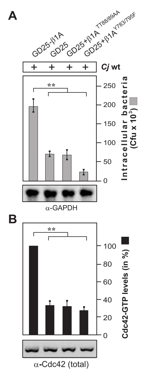
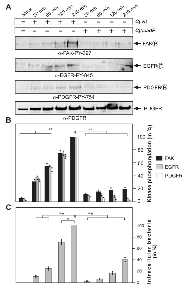
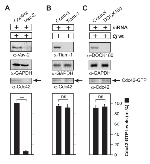
Results: Using high-resolution scanning electron microscopy, GTPase pulldowns, G-Lisa and gentamicin protection assays we found that each studied host factor is necessary for induction of Cdc42-GTP and efficient invasion. Interestingly, filopodia formation and associated membrane dynamics linked to invasion were only seen during infection of wild-type but not in knockout cells. Infection of cells stably expressing integrin-beta1 variants with well-known defects in fibronectin fibril formation or FAK signaling also exhibited severe deficiencies in Cdc42 activation and bacterial invasion. We further demonstrated that infection of wild-type cells induces increasing amounts of phosphorylated FAK and growth factor receptors (EGFR and PDGFR) during the course of infection, correlating with accumulating Cdc42-GTP levels and C. jejuni invasion over time. In studies using pharmacological inhibitors, silencing RNA (siRNA) and dominant-negative expression constructs, EGFR, PDGFR and PI3-kinase appeared to represent other crucial components upstream of Cdc42 and invasion. siRNA and the use of Vav1/2-/- knockout cells further showed that the guanine exchange factor Vav2 is required for Cdc42 activation and maximal bacterial invasion. Overexpression of certain mutant constructs indicated that Vav2 is a linker molecule between Cdc42 and activated EGFR/PDGFR/PI3-kinase. Using C. jejuni mutant strains we further demonstrated that the fibronectin-binding protein CadF and intact flagella are involved in Cdc42-GTP induction, indicating that the bacteria may directly target the fibronectin/integrin complex for inducing signaling leading to its host cell entry.

Conclusion: Collectively, our findings led us propose that C. jejuni infection triggers a novel fibronectin→integrin-beta1→FAK/Src→EGFR/PDGFR→PI3-kinase→Vav2 signaling cascade, which plays a crucial role for Cdc42 GTPase activity associated with filopodia formation and enhances bacterial invasion.


Figures

Figure 1
Figure 1
C. jejuni-triggered Cdc42 activation is time-dependent and requires intact lipid rafts. (A) Quantification of Cdc42 activity during the course of infection. INT-407 cells were infected with wt C. jejuni 81-176 for indicated periods of time. The presence of active Cdc42-GTP was quantified by G-Lisa and GST-CRIB pulldown. One hundred % of GTPase activity corresponds to the highest amount of detected Cdc42-GTP level (right lane). Similar quantities of total Cdc42 and GAPDH were confirmed by Western blotting. (B) Effect of Cdc42 expression knockdown on C. jejuni invasion. INT-407 cells were transfected with Cdc42-siRNA as well as a scrambled siRNA as control. After 48 hours, cells were infected with C. jejuni for 6 hours. Intracellular bacteria were quantified by gentamicin protection assays. Immunoblotting with α-Cdc42 antibody confirmed down-regulation of the protein. GAPDH expression levels were determined as control. (C) Effects of MβCD targeting lipid rafts on host cell internalization of C. jejuni. INT-407 monolayers were pre-incubated with the indicated concentrations of MβCD for 30 min, followed by 6 hours infection with wt C. jejuni 84-25. Intracellular C. jejuni were quantified by gentamicin protection assays. The presence of active Cdc42-GTP was analyzed by CRIB-GST pulldown and quantified. One hundred % of activity corresponds to the highest amount of detected Cdc42-GTP level (lane 2). Similar quantities of total Cdc42 and GAPDH were confirmed by Western blotting. (*) P ≤ 0.05 and (**) P ≤ 0.005 were considered as statistically significant as compared to the control.
Figure 2
Figure 2
Importance of fibronectin, integrin-β1, FAK and Src kinases expression on C. jejuni invasion. The following cells lines were infected with wt C. jejuni 81-176 for 6 hours. (A) Fibronectin-deficient cells (Fn-/-) and corresponding floxed wt cells (Fn+/+), (B) integrin-β1-deficient cells (GD25) and GD25 stably re-expressing wt integrin-β1A (GD25-β1A) cells, (C) FAK-deficient cells (FAK-/-) and FAK-/- cells stably re-expressing wt FAK and (D) Src kinase-deficient cells (SYF-/-) and SYF-/- cells stably re-expressing wt c-src. Intracellular C. jejuni were quantified by gentamicin protection assays, and Cdc42 activation by CRIB-GST pulldowns. (**) P ≤ 0.005 was considered as statistically significant. Fibronectin, integrin-β1, FAK and Src expression was analyzed by immunoblotting. GAPDH expression levels were determined as loading control.
Figure 3
Figure 3
C. jejuni invasion is impaired in cells expressing integrin-β1 point mutations exhibiting defects in fibronectin fibril organisation or FAK phosphorylation. (A) Integrin-β1-deficient cells (GD25) and GD25 stably re-expressing mutated integrin subunit β1A (GD25-β1 ATT788-9AA or GD25-β1 AY783/795F or wild-type β1A (GD25-β1A) cells were infected with wt C. jejuni 81-176 for 6 hours. Intracellular C. jejuni were quantified by gentamicin protection assays. (B) The presence of active Cdc42-GTP was quantified by CRIB-GST pulldowns. One hundred % of activity corresponds to the highest amount of detected Cdc42-GTP level. (**) P ≤ 0.005 was considered as statistically significant. Similar quantities of total Cdc42 and GAPDH were confirmed by Western blotting.
Figure 4
Figure 4
High resolution FESEM of C. jejuni-induced filopodia formation. Representative sections of wild-type fibroblasts incubated for 6 hours with wt C. jejuni 81-176 (A) and non-infected fibroblast control cells that were mock-treated (B) are shown. Infection revealed the occurrence of membrane protrusion events with long filopodia at the periphery and on top of cells which were only sporadically seen in the non-infected control cells (blue arrows).
Figure 5
Figure 5
High resolution FESEM of C. jejuni-induced filopodia formation and invasion. (A) Infection of GD25 knockout cells with wt C. jejuni 81-176 (yellow arrows) for 6 hours revealed bacterial attachment to the cell surface with short microspikes (green arrowheads) present, but membrane dynamics events or invasion were rarely seen. Similar observations were made with infected fibronectin-/- or FAK-/- cells. (B) Infecting C. jejuni in wt cells were regularly associated with long filopodia (blue arrows) membrane ruffling (red arrows), as well as elongated microspikes (green arrowheads).
Figure 6
Figure 6
Importance of CadF for C. jejuni-induced FAK, EGFR and PDGFR activation. (A) FAK-positive fibroblasts were infected with wt C. jejuni F38011 or isogenic F38011ΔcadF for indicated periods of time. FAK, EGFR or PDGFR activation was analysed by immunoblotting with indicated antibodies. Total PDGFR expression levels were determined as loading control. (B) Quantification of FAK, EGFR and PDGFR kinase phosphorylation during the course of infection. One hundred % of activity corresponds to the highest amount of phosphorylation detected per experiment and selected kinase (lane 5). (C) Intracellular C. jejuni were quantified by gentamicin protection assays. (*) P ≤ 0.005 and (**) P ≤ 0.005 were considered as statistically significant.
Figure 7
Figure 7
Importance of the CadF and flagellar apparatus for C. jejuni-induced activation of Cdc42 and bacterial invasion. (A) FAK+/+ and FAK-/- cells were infected with wt C. jejuni F38011 or isogenic F38011ΔcadF for the indicated periods of time. Quantification of Cdc42-GTP levels by CRIB-GST pulldown during the course of infection. One hundred % of activity corresponds to the highest amount of detected Cdc42-GTP level (lane 4). (B) FAK-positive cells were infected with the indicated strains in a time-course. The presence of bound, active Cdc42-GTP was analyzed in CRIB-GST pulldown assays followed by Western blotting using α-Cdc42 antibody. Similar quantities of individual GTPases at every time point were confirmed by Western blotting using equivalent volumes of cell lysates. (C) Quantification of Cdc42-GTP levels during the course of infection. One hundred % of activity corresponds to the highest amount of detected Cdc42-GTP level (lane 3). The amount of intracellular bacteria was quantified by gentamicin protection assays under the same experimental conditions. (**) P ≤ 0.005 were considered as statistically significant as compared to the control.
Figure 8
Figure 8
Importance of guanine exchange factor Vav2 for C. jejuni-induced Cdc42 activation. INT-407 cells were transfected for 48 hours with siRNA for Vav2 (A), Tiam-1 (B) or DOCK180 (C) as well as a scrambled siRNA as control. Immunoblotting with the indicated antibodies confirmed knockdown of the respective proteins. GAPDH expression levels were determined as control. Quantification of Cdc42 GTPase activity after infection with wt C. jejuni 81-176 for 6 hours. The presence of bound, active Cdc42-GTP was analyzed in CRIB-GST pulldown assays followed by Western blotting using α-Cdc42 antibody. One hundred % of activity corresponds to the highest amount of detected Cdc42-GTP level (lane 1).
Figure 9
Figure 9
Downregulation, elimination or interference with important Vav2 functions reduces the uptake of C. jejuni in host cells. (A) INT-407 cells were transfected with siRNA against Vav2 or a scrambled siRNA as control. After 48 hours, cells were infected with wt C. jejuni 81-176 for 6 hours. Intracellular bacteria were quantified by gentamicin protection assays. (B) The presence of active Rac1-GTP and Cdc42-GTP was quantified by CRIB-GST pulldowns. One hundred % of activity corresponds to the highest amount of detected GTPase-GTP level. (C) INT-407 cells were transfected with indicated Myc-tagged or (D) GFP-tagged Vav2 constructs. After 48 hours, cells were infected with wt C. jejuni 81-176 for 6 hours. Intracellular bacteria were quantified by gentamicin protection assays. Expression of the individual Vav2 constructs was verified by Western blot analysis. GAPDH expression levels were determined as control. (E) Vav2-deficient cells (Vav1/2-/-) or Vav2-expressing control fibroblasts (Vav1/2+/+) were infected for 6 hours with C. jejuni. Intracellular and cell-associated bacteria were quantified by gentamicin protection assays. (*) P ≤ 0.05 and (**) P ≤ 0.005 were considered as statistically significant.
Figure 10
Figure 10
Importance of FAK, EGFR, PDGFR and PI3-kinase activities for C. jejuni-induced activation of Cdc42 and bacterial invasion. (A) INT-407 monolayers were pre-incubated for 30 min with the indicated pharmacological inhibitors and infected with C. jejuni for 6 hours. Intracellular C. jejuni were quantified by gentamicin protection assays. The presence of active Cdc42-GTP was quantified by CRIB-GST pulldowns. One hundred % of activity corresponds to the highest amount of detected Cdc42-GTP level (lane 2). (B) Effect of overexpression of dominant-negative forms of PDGFR and EGFR on C. jejuni uptake. 48 hours post transfection INT-407 cells were infected with C. jejuni for 6 hours. Intracellular bacteria were quantified by gentamicin protection assays. Expression of the individual constructs was verified by Western blotting. GAPDH expression levels were determined as control. (*) P ≤ 0.05 and (**) P ≤ 0.005 were considered as statistically significant.
Figure 11
Figure 11
Model for C. jejuni-induced signaling leading to Cdc42 activation and bacterial invasion. C. jejuni adheres to host cells via the fibronectin-binding protein CadF, which acts as a bridge engaging the integrin-β1 receptor. Integrin occupancy and clustering in lipid rafts leads to recruitment and activation of the non-receptor tyrosine kinase FAK. Phosphorylation of FAK and Src triggers a cascade of signals resulting in the formation of protein complexes leading to activation of other signaling factors as indicated. Assembly of integrin-dependent signal complexes leads to phosphorylation and transactivation of PDGFR and EGFR, followed by stimulation of PI3-K and Vav2. Activated Vav2 then induces the activation of Cdc42. This signaling potentially causes localized actin and/or microtubule rearrangements at the site of C. jejuni entry, resulting in bacterial uptake. In addition to CadF, the C. jejuni flagellum also appears to play a role in the described signal cascades. If the flagellum participates by sole bacterial motility, by translocating bacterial effector proteins or targeting a host receptor directly is not yet clear and needs to be investigated in future studies.

Similar articles

Cited by

References

    1. World Health Organization. Global burden of disease (GBD) 2002 estimates. WHO. Geneva, Switzerland; 2004. http://www.who.int/topics/global_burden_of_disease/en/
    1. Nachamkin I, Szymanski CM, Blaser MJ. Campylobacter. Washington, DC: ASM Press; 2008.
    1. Oyarzabal OA, Backert S. Microbial Food Safety: An Introduction. Heidelberg (Germany): Springer Verlag; 2012. in press .
    1. Young KT, Davis LM, DiRita VJ. Campylobacter jejuni: molecular biology and pathogenesis. Nat Rev Microbiol. 2007;5:665–679. doi: 10.1038/nrmicro1718. - DOI - PubMed
    1. Blaser MJ, Engberg J. In: Campylobacter. Nachamkin I, Szymanski CM, Blaser MJ, editor. Washington, DC: ASM Press; 2008. pp. 99–121.