. 2011 Dec 29:8:562.
doi: 10.1186/1743-422X-8-562.

Orf virus DNA vaccines expressing ORFV 011 and ORFV 059 chimeric protein enhances immunogenicity

Affiliations

Orf virus DNA vaccines expressing ORFV 011 and ORFV 059 chimeric protein enhances immunogenicity

Kui Zhao et al. Virol J. .

Abstract

Background: ORFV attenuated live vaccines have been the main prophylactic measure against contagious ecthyma in sheep and goats in the last decades, which play an important role in preventing the outbreak of the disease. However, the available vaccines do not induce lasting immunity in sheep and goats. On the other hand, variation in the terminal genome of Orf virus vaccine strains during cell culture adaptation may affect the efficacy of a vaccine. Currently, there are no more effective antiviral treatments available for contagious ecthyma.

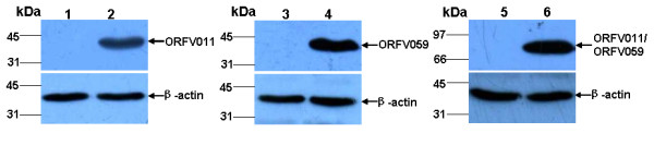
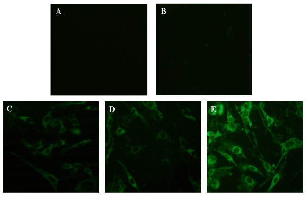
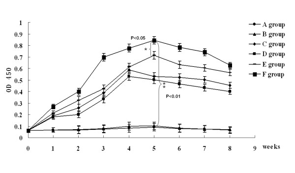
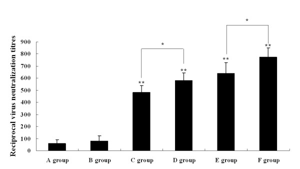
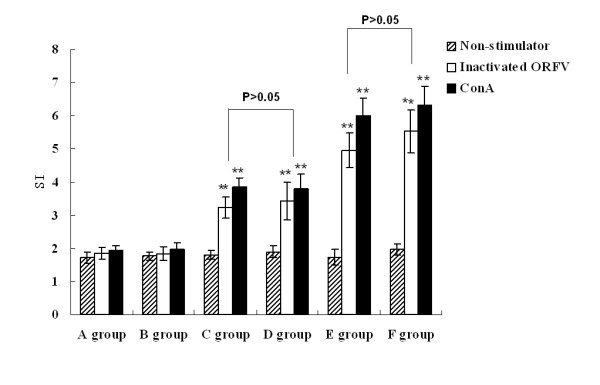
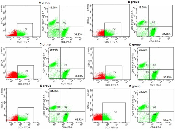
Results: We constructed three eukaryotic expression vectors pcDNA3.1-ORFV011, pcDNA3.1-ORFV059 and pcDNA3.1-ORFV011/ORFV059 and tested their immunogenicity in mouse model. High level expression of the recombinant proteins ORFV011, ORFV059 and ORFV011/ORFV059 was confirmed by western blotting analysis and indirect fluorescence antibody (IFA) tests. The ORFV-specific antibody titers and serum IgG1/IgG2a titers, the proliferation of lymphocytes and ORFV-specific cytokines (IL-2, IL-4, IL-6, IFN-γ, and TNF-α) were examined to evaluate the immune responses of the vaccinated mice. We found that mice inoculated with pcDNA3.1-ORFV 011/ORFV059 had significantly stronger immunological responses than those inoculated with pcDNA3.1-ORFV011, pcDNA3.1-ORFV059, or pcDNA3.1-ORFV011 plus pcDNA3.1-ORFV059. Compared to other vaccine plasmids immunized groups, pcDNA3.1-ORFV011/ORFV059 immunized group enhances immunogenicity.

Conclusions: We concluded that DNA vaccine pcDNA3.1-ORFV011/ORFV059 expressing ORFV011 and ORFV059 chemeric-proteins can significantly improve the potency of DNA vaccination and could be served as more effective and safe approach for new vaccines against ORFV.


Figures

Figure 1
Figure 1
Western blot analysis of ORFV011 protein, ORFV059 protein and ORFV 011/ORFV 059 chemeric-proteins. OFTu cells were transfected with pcDNA 3.1(+) plasmid (control) or pcDNA 3.1-ORFV 011, pcDNA 3.1-ORFV 059, pcDNA 3.1-ORFV 011/ORFV 059 vaccine plasmids, respectively. After 48 h post-transfection, lysates collected from different vaccine plasmids-transfected OFTu cell were examined by SDS-PAGE -Western blotting with rabbit anti-ORFV polyclonal antibody (kindly provided by the Jilin Institute for Veterinary Research, diluted at 1:100). ORFV011 protein, ORFV059 protein and ORFV 011/ORFV059 chimeric-proteins bands could be detected in pcDNA3.1-ORFV011, pcDNA3.1-ORFV059 and pcDNA3.1-ORFV011/ORFV059 recombinant vaccine plasmids-transfected OFTu cell lysates (Figure 1, Lanes 2, 4, 6), but no corresponding protein bands were detected in the pcDNA3.1(+)-transfected cell lysates (Figure 1, Lanes 1, 3, 5). Lanes 2, 4, 6: OFTu cells transfected with pcDNA 3.1-ORFV011, pcDNA 3.1-ORFV059 or pcDNA 3.1-ORFV011/ORFV059 vaccine plasmids, respectively. Lanes 1, 3, 5: OFTu cells transfected with pcDNA 3.1(+) plasmid (control).
Figure 2
Figure 2
The intracellular location of ORFV011 protein, ORFV059 protein and ORFV 011/ORFV059 chemeric-proteins in transfected OFTu cells. The OFTu cells were seeded in 6-well culture plates and un-transfected (Figure 2a) or transfected with pcDNA3.1(+) vector (Figure 2b), pcDNA3.1-ORFV011 plasmid (Figure 2c), pcDNA3.1-ORFV059 plasmid (Figure 2d) or pcDNA3.1-ORFV011/ORFV059 plasmid (Figure 2e) for 48 h. After 48 h post-transfection, the cells were fixed with 80% acetone for 10 min at -20°C, rehydrated in PBS, labeled with a rabbit anti-ORFV polyclonal antibody, and washed three times with PBS. FITC-conjugated goat anti-rabbit IgG (H + L) (1:500 dilution in PBS) was added to the OFTu cell mixtures for 30 min at room temperature, and the cells were washed and observed with a fluorescence microscopy. Magnification: 400 ×.
Figure 3
Figure 3
Group mean optical density ratios and standard errors for specific anti-ORFV-IgG antibody responses in the serum samples of immunized mice of different groups. The sera were collected from mice in groups (pcDNA3.1-ORFV011 group, pcDNA3.1-ORFV059 group, pcDNA3.1-ORFV011/ORFV059 group, pcDNA3.1-ORFV 011 plus pcDNA3.1-ORFV059 group, pcDNA3.1(+) group and PBS group) at weeks 0, 1, 2, 3, 4, 5, 6, 7, 8 and were inactivated at 56° for 30 min. The ORFV-specific antibody responses were determined using an indirect ELISA, with the purified ORFV as the coating antigen. As shown in Figure 3, the levels of anti-ORFV-IgG antibodies were developed in different DNA vaccine plasmid- immunized group. One week after the final boost immunization, the level of anti-ORFV-IgG antibodies was higher in the pcDNA3.1-ORFV 011/ORFV 059 immunized group than any of the other groups tested (OD450 value: 0.829 ± 0.014, **P < 0.01). The levels of anti-ORFV-IgG antibodies in both of pcDNA3.1-ORFV 011 group and pcDNA3.1-ORFV 059 group were very nearly, but in the pcDNA3.1-ORFV 011 group increased slightly compared with the pcDNA3.1-ORFV 059 group (OD450 value: 0.579 ± 0.009 vs 0.518 ± 0.003, P > 0.05). The levels of anti-ORFV-IgG antibodies was higher in the pcDNA3.1-ORFV 011/ORFV 059 group than pcDNA3.1-ORFV 011 plus pcDNA3.1-ORFV 059 group (OD450 value: 0.829 ± 0.014 vs 0.687 ± 0.024, *P < 0.05). A group: PBS; B group: pcDNA3.1(+) vector; C group: pcDNA3.1-ORFV011; D group: pcDNA3.1-ORFV059; E group: pcDNA3.1-ORFV011 plus pcDNA3.1-ORFV059; F group: pcDNA3.1-ORFV011/ORFV059.
Figure 4
Figure 4
The subclasses IgG1 and IgG2a of anti-ORFV IgG antibodies in the serum samples of immunized mice of different groups. Sera were taken from immunized mice of different groups 1 week post the final boost immunization. The titration of ORFV-specific immunoglobulin (IgG) subclasses 1 and 2a was detected by ELISA. As shown in Figure 4, the assay of IgG2a and IgG1 antibodies performed at 1 week post the final boost immunization indicates a significant difference between vaccine plasmids groups (including pcDNA 3.1-ORFV011 group, pcDNA 3.1-ORFV059 group, pcDNA3.1-ORFV011 plus pcDNA3.1-ORFV059 group and pcDNA3.1-ORFV011/ORFV059 group) and control groups (including PBS and pcDNA3.1(+) group) (**P < 0.01). A group: PBS; B group: pcDNA3.1(+) vector; C group: pcDNA3.1-ORFV011; D group: pcDNA3.1-ORFV059; E group: pcDNA3.1-ORFV011 plus pcDNA3.1-ORFV059; F group: pcDNA3.1-ORFV011/ORFV059.
Figure 5
Figure 5
Virus-neutralizing titer assay. The virus-neutralizing titers were determined in sera pooled from immunized mice of different groups 2 weeks post immunization. The sera (diluted 1:5 in MEM medium) were heat-activated for 30 min at 56°C, then serially diluted two-fold in 96-well flat bottom plates and incubated with 0.1 μg of purified ORFV diluted in 50 μl MEM for 90 min at 37°C. All sera were run in triplicate. Subsequently, 100 μl of OFTu cell suspension were added to each well at a concentration of 45,000 cells/well. The plates were incubated for 5 days at 37°C in a 5% CO2 incubator. The titer is expressed as ND50 and calculated using the Reed and Muench method. As shown in Figure 5, the assay of virus-neutralizing titers performed at 2 weeks post immunization indicates a significant difference between four vaccine plasmids groups (including pcDNA 3.1-ORFV 011 group, pcDNA 3.1-ORFV 059 group, pcDNA-ORFV 011 plus pcDNA-ORFV 059 group and pcDNA-ORFV 011/ORFV 059 group) and control groups (including PBS and pcDNA3.1(+) group) (**P < 0.01). Among the four vaccine plasmids groups, the virus-neutralizing titers in the pcDNA-ORFV 011/ORFV 059 group was significantly higher than that of pcDNA-ORFV 011 plus pcDNA-ORFV 059 group (*P < 0.05). A group: PBS; B group: pcDNA3.1(+) vector; C group: pcDNA3.1-ORFV011; D group: pcDNA3.1-ORFV059; E group: pcDNA3.1-ORFV011 plus pcDNA3.1-ORFV059; F group: pcDNA3.1-ORFV011/ORFV059.
Figure 6
Figure 6
Proliferation of spleen lymphocytes in the different groups. Six groups of 6-8-week-old female Balb/c mice were immunized by skin scarification with one of the following formulations: (A) PBS; (B) pcDNA3.1(+) vector; (C) pcDNA3.1-ORFV011; (D) pcDNA3.1-ORFV059; (E) pcDNA3.1-ORFV011 plus pcDNA3.1-ORFV059; (F) pcDNA3.1-ORFV011/ORFV059. Each bar represents the group mean (N = 10) ± S.E.M. of SI determined in triplicate. The mitogen, Con A served as a positive antigen and positive control. As shown in Figure 5, mice immunized with pcDNA3.1-ORFV011/ORFV059 (F group) induced a stronger proliferation response compared with background OD (0.281 ± 0.123) (**P < 0.01). The E group, immunized with pcDNA3.1-ORFV011 plus pcDNA3.1-ORFV059, also resulted displayed a higher stimulation index (SI) than mice immunized with PBS, pcDNA3.1 (+) or vaccine plasmid alone. However, the stimulation index (SI) in pcDNA3.1-ORFV011/ORFV059 group increased slightly compared with the pcDNA3.1-ORFV011 plus pcDNA3.1-ORFV059 group (P > 0.05), and the stimulation index (SI) were no obvious difference between pcDNA3.1-ORFV 011 group and pcDNA3.1-ORFV 059 group (P > 0.05).
Figure 7
Figure 7
The CD4+ and CD8+ T lymphocyte subsets in the different groups were assessed by FACS analysis at day 10 post the final boost immunization. Spleen lymphocytes of mice in different immunized groups were repectively enriched from the spleens at days 10 post the final boost immunization and cultured in RPMI 1640 medium. The CD4+ and CD8+ T lymphocyte subsets in the different groups were assessed by FACS in order to examine whether or not DNA vaccine constructs immunized mice by skin scarification can induce a T-cell response in mice and further activate cell immunity response. (A) PBS; (B) pcDNA3.1(+) vector; (C) pcDNA3.1-ORFV011; (D) pcDNA3.1-ORFV059; (E) pcDNA3.1-ORFV011 plus pcDNA3.1-ORFV059; (F) pcDNA3.1-ORFV011/ORFV059.
Figure 8
Figure 8
The production of ORFV-specific cytokines in the supernatant of splenocytes stimulated with purified ORFV at day 10 post the final boost immunization. Six groups of of 6-8-week-old female Balb/c mice were immunized by skin scarification with one of the following formulations: (A) PBS; (B) pcDNA3.1(+) vector; (C) pcDNA3.1-ORFV011; (D) pcDNA3.1-ORFV059; (E) pcDNA3.1-ORFV011 plus pcDNA3.1-ORFV059; (F) pcDNA3.1-ORFV011/ORFV059. The levels of cytokines production (IFN-γ, TNF-α, IL-2, IL-4 and IL-6) in the supernatant of splenocytes stimulated with purified ORFV were determined by ELISA assay. Each bar represents the group mean (N = 10) ± S.E.M. of cytokines levels determined in triplicate.

Similar articles

Cited by

References

    1. Haig DM, Mercer AA. Ovine diseases. Orf. Vet Res. 1998;29:311–326. - PubMed
    1. De la Concha-Bermejillo A. In: Health Hazards in Veterinary Practice. 3. Farris R, Mahlow J, Newman E, Nix B, editor. American Veterinary Medical Association, Schaumburg; 1995. Poxviral diseases; pp. 55–56.
    1. Gumbrell RC, McGregor DA. Outbreak of severe fatal orf in lambs. Vet Rec. 1997;141:150–151. doi: 10.1136/vr.141.6.150. - DOI - PubMed
    1. Mazur C, Machado RD. Detection of contagious pustular dermatitis virus of goats in a severe outbreak. Vet Rec. 1989;125:419–420. doi: 10.1136/vr.125.16.419. - DOI - PubMed
    1. Mazur C, Machado RD. The isolation and identification of the contagious ecthyma virus of caprines in cell cultures. Rev Microbiol São Paulo. 1990;21:127–130. - PubMed

Publication types

MeSH terms

LinkOut - more resources