. 2011 Dec 28:11:268.
doi: 10.1186/1471-2180-11-268.

The promoter of filamentation (POF1) protein from Saccharomyces cerevisiae is an ATPase involved in the protein quality control process

Affiliations

The promoter of filamentation (POF1) protein from Saccharomyces cerevisiae is an ATPase involved in the protein quality control process

Iris M Costa et al. BMC Microbiol. .

Abstract

Background: The gene YCL047C, which has been renamed promoter of filamentation gene (POF1), has recently been described as a cell component involved in yeast filamentous growth. The objective of this work is to understand the molecular and biological function of this gene.

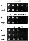
Results: Here, we report that the protein encoded by the POF1 gene, Pof1p, is an ATPase that may be part of the Saccharomyces cerevisiae protein quality control pathway. According to the results, Δpof1 cells showed increased sensitivity to hydrogen peroxide, tert-butyl hydroperoxide, heat shock and protein unfolding agents, such as dithiothreitol and tunicamycin. Besides, the overexpression of POF1 suppressed the sensitivity of Δpct1, a strain that lacks a gene that encodes a phosphocholine cytidylyltransferase, to heat shock. In vitro analysis showed, however, that the purified Pof1p enzyme had no cytidylyltransferase activity but does have ATPase activity, with catalytic efficiency comparable to other ATPases involved in endoplasmic reticulum-associated degradation of proteins (ERAD). Supporting these findings, co-immunoprecipitation experiments showed a physical interaction between Pof1p and Ubc7p (an ubiquitin conjugating enzyme) in vivo.

Conclusions: Taken together, the results strongly suggest that the biological function of Pof1p is related to the regulation of protein degradation.


Figures
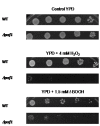
Figure 1
Figure 1
Δpof1 cells are sensitive to oxidative stress. A representative viability assay showing cells exposed to hydrogen peroxide (H2O2) or tert-butyl hydroperoxide (t-BOOH) on rich solid media (YPD). The cells (collected at stationary phase) were diluted to OD600 nm = 0.2, followed by 4 serial dilutions of 5X. A total of 5 μL of each dilution were spotted on the plates, which were incubated at 30°C for 48 h and photographed.
Figure 2
Figure 2
POF1 and PCT1 sequences and functional analyses. (A) Clustal W (Megalign software) primary sequence alignment of the cytidylyltransferase family. The conserved motif HxxH is enclosed in the box. Ct human = choline cytidylyltransferase from humans (gi 166214967); et human = ethanolamine cytidylyltransferase from humans (gi 1817548); pct1 yeast = phosphocholine cytidylyltransferase from S. cerevisiae (gi 1323361); ycl047c = Pof1p (gi 6319802). (B) Complementation assays. The cells were exposed to heat shock (37°C) or control conditions (30°C) for 17 h, serially diluted and spotted on YPD plates, which were incubated for 48 h at 30°C and photographed. The asterisk denotes cells transformed with the plasmid pYES-TOPO+POF1 for overexpression of Pof1.
Figure 3
Figure 3
Pof1p purification and activity analyses. (A) SDS-PAGE showing the purification of recombinant Pof1p obtained through metal affinity chromatography. Lane 1: molecular weight standard; subsequent lanes were different fractions obtained during the elution process. (B) Thin layer chromatography analyses to observe Pof1p ATP transferase activity; the controls were included to assay for alterations in CTP and ATP. See the Materials and Methods section for details.
Figure 4
Figure 4
Δpof1 cells are sensitive to protein unfolding. The cells from the stationary phase of growth were diluted to OD600 nm = 0.2, followed by 4 serial dilutions of 5X. A total of 5 μL of each dilution were spotted on SD complete media plates containing no unfolding agent, DTT or tunicamycin. The plates were incubated at 30°C for 48 h and photographed.
Figure 5
Figure 5
ATPase activity of Pof1p and its physical interaction with Ubc7 (ubiquitin conjugase 7) protein. (A) Hydrolysis of ATP as measured by the PiPer™ Phosphate Assay Kit (Invitrogen). (-) ATP represents the result of the reaction in the absence of ATP; (-) POF1 represents the result of the spontaneous ATP hydrolysis in the absence of Pof1p; the lane labeled "Pof1p + ATP" contains the complete reaction. The assays were performed at 37°C for 1 h. (B) Western blot analyses of Ubc7p using the commercial antibody Ube2G2 (Abcam). The fractions were obtained from co-immunoprecipitation assays using Pof1p polyclonal antibody from the following protein extract: WT = wild type; Δpct1 and Δpof1. The asterisk shows the Pof1p-Ubc7p complex. CE = total soluble wild type cell extract; IgG LC = IgG light chain. The arrow points Ubc7p dimer.
Figure 6
Figure 6
Immunocytochemistry assays to study Pof1 protein cell localization and distribution. The POF1 null cells were used as a negative control to establish antibody background levels.

References

    1. Leidhold C, Voos W. Chaperones and proteases--guardians of protein integrity in eukaryotic organelles. Ann N Y Acad Sci. 2007. pp. 72–86. - PubMed
    1. Carvalho P, Goder V, Rapoport TA. Distinct ubiquitin-ligase complexes define convergent pathways for the degradation of ER proteins. Cell. 2006;126:361–373. doi: 10.1016/j.cell.2006.05.043. - DOI - PubMed
    1. Denic V, Quan EM, Weissman JS. A luminal surveillance complex that selects misfolded glycoproteins for ER-associated degradation. Cell. 2006;126:349–359. doi: 10.1016/j.cell.2006.05.045. - DOI - PubMed
    1. Carvalho P, Stanley AM, Rapoport TA. Retrotranslocation of a misfolded luminal ER protein by ubiquitin-ligase Hrd1p. Cell. 2010;143:579–591. doi: 10.1016/j.cell.2010.10.028. - DOI - PMC - PubMed
    1. Turner GC, Varshavsky A. Detecting and measuring cotranslational protein degradation in vivo. Science. 2000;289:2117–2120. doi: 10.1126/science.289.5487.2117. - DOI - PubMed

Publication types

MeSH terms

Substances