Review
. 2011 Dec;17(38):4294-307.
doi: 10.2174/138161211798999438.

Cyclotides, a novel ultrastable polypeptide scaffold for drug discovery

Affiliations
Review

Cyclotides, a novel ultrastable polypeptide scaffold for drug discovery

Andrew Gould et al. Curr Pharm Des. 2011 Dec.

Abstract

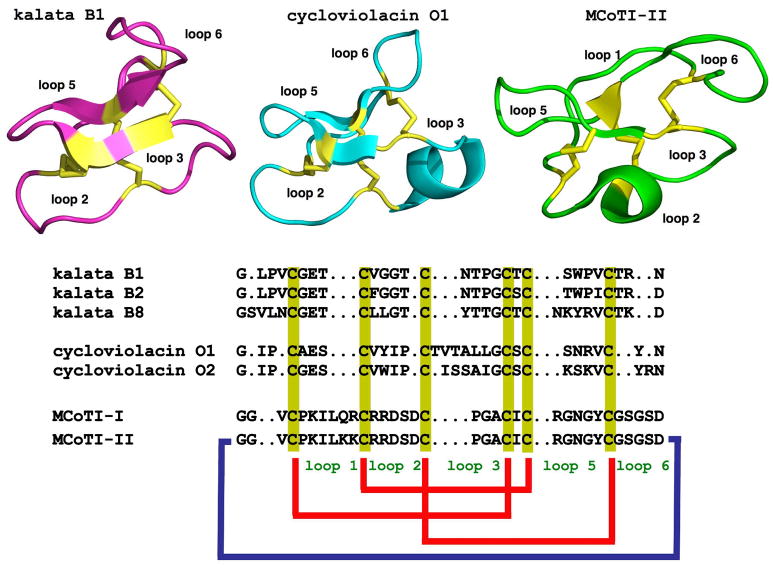
Cyclotides are a unique and growing family of backbone cyclized peptides that also contain a cystine knot motif built from six conserved cysteine residues. This unique circular backbone topology and knotted arrangement of three disulfide bonds makes them exceptionally stable to thermal, chemical, and enzymatic degradation compared to other peptides of similar size. Aside from the conserved residues forming the cystine knot, cyclotides have been shown to have high variability in their sequences. Consisting of over 160 known members, cyclotides have many biological activities, ranging from anti-HIV, antimicrobial, hemolytic, and uterotonic capabilities; additionally, some cyclotides have been shown to have cell penetrating properties. Originally discovered and isolated from plants, cyclotides can also be produced synthetically and recombinantly. The high sequence variability, stability, and cell penetrating properties of cyclotides make them potential scaffolds to be used to graft known active peptides or engineer peptide-based drug design. The present review reports recent findings in the biological diversity and therapeutic potential of natural and engineered cyclotides.


Figures

Figure 1
Figure 1
Primary and tertiary structures of cyclotides from the Möbius (kalata B1, pdb code: 1NB1), bracelet (cycloviolacin O1, pdb code: 1NBJ) and trypsin inhibitor (MCoTI-II, pdb code: 1IB9) subfamilies. The sequence of kalata B8, a novel hybrid cyclotide isolated from the plant O. affinis is also shown. Conserved cysteine residues are marked in yellow and disulfide connectivities in red. The circular backbone topology is shown with a blue line. Figure adapted from Ref. [4].
Figure 2
Figure 2
General features of the cyclic cystine knot (CCK) topology found in cyclotides. A. Detailed structure the cystine knot core and the connecting loops. The six Cys residues are labeled I through VI whereas loops connecting the different Cys residues are designated as loop 1 through 6, in numerical order from the N- to the C-terminus. B. Möbius (right) and bracelet (left) cyclotides are defined by the presence (Möbius) or absence (bracelet) of a Pro residue in loop 5 that introduces a twist in the circular backbone topology.
Figure 3
Figure 3
Genetic origin and biosynthesis of cyclotides in plants. A. Rubiacea and Violaceae plants have dedicated genes for the production of cyclotides [13]. These cyclotide precursors comprise an ER signal peptide, an N-terminal Pro region, the N-terminal repeat (NTR), the mature cyclotide domain and a C-terminal flanking region (CTR). Cyclotides from the Fabaceae family of plants isolated recently from C. ternatea [9, 10], show an ER signal peptide immediately followed by the cyclotide domain, which is flanked at the C-terminus by a peptide linker and the albumin a-chain. In this case, the cyclotide domain replaces albumin-1 b-chain. The genetic origin of the trypsin inhibitor cyclotides MCoTI-I/II (found in the seeds of M. cochinchinensis) remains yet to be determined. B. Scheme representing the proposed mechanism of protease-catalyzed cyclotide cyclization. It has been proposed that the cyclization step is mediated by an asparaginyl endopeptidase (AEP), a common Cys protease found in plants. The cyclization takes place at the same time as the cleavage of the C-terminal pro-peptide from the cyclotide precursor protein through a transpeptidation reaction [66]. The transpeptidation reaction involves an acyl-transfer step from the acyl-AEP intermediate to the N-terminal residue of the cyclotide domain [67]. Figure adapted from Refs. [4, 11].
Figure 4
Figure 4
Chemical synthesis of cyclotides by means of an intramolecular native chemical ligation (NCL). This approach requires the chemical synthesis of a linear precursor bearing an N-terminal Cys residue and an α-thioester moiety at the C-terminus. The linear precursor can be cyclized first under reductive conditions and then folded using a redox buffer containing reduced and oxidized gluthathione (GSH). Alternatively, the cyclization and folding can be efficiently accomplished in a single pot reaction when the cyclization is carried out in the presence of reduced GSH as the thiol cofactor [18, 20].
Figure 5
Figure 5
Biosynthetic approach for the recombinant expression of cyclotides using E. coli expression systems. Cleavage of the leading signal either in vitro [18] or in vivo [19, 20] by appropriate proteases provides the N-terminal Cys residue required for the cyclization. The backbone cyclization of the linear precursor is then mediated by a modified protein splicing unit or intein. Once the linear precursor is cyclized, folding is spontaneous for kalata B1 and MCoTI-I/II cyclotides [–20].
Figure 6
Figure 6
Summary of some of the modifications made to kalata B1 and MCoTI-II to elicit novel biological activities. Modifications include the grafting of peptides onto various loops of kalata B1 [75] and MCoTI-II [17, 134, 135]. Cyclotide based libraries have been also generated using loops 1, 2, 3, 4 and 5 of MCoTI-I to study the effect of individual mutations on the biological activity and folding ability of MCoTI-I mutants [20]. The locations of the changes introduced into the cyclotide framework are illustrated using the MCoTI-II structure (pdb ID: 1IB9).

Similar articles

Cited by

References

    1. Daly NL, Rosengren KJ, Craik DJ. Discovery, structure and biological activities of cyclotides. Adv Drug Deliv Rev. 2009;61:918–930. - PubMed
    1. Puttamadappa SS, Jagadish K, Shekhtman A, Camarero JA. Backbone Dynamics of Cyclotide MCoTI-I Free and Complexed with Trypsin. Angew Chem Int Ed Engl. 2010;49:7030–7034. - PMC - PubMed
    1. Colgrave ML, Craik DJ. Thermal, chemical, and enzymatic stability of the cyclotide kalata B1: the importance of the cyclic cystine knot. Biochemistry. 2004;43:5965–5975. - PubMed
    1. Garcia AE, Camarero JA. Biological activities of natural and engineered cyclotides, a novel molecular scaffold for peptide-based therapeutics. Curr Mol Pharmacol. 2010;3:153–163. - PMC - PubMed
    1. Saether O, Craik DJ, Campbell ID, Sletten K, Juul J, Norman DG. Elucidation of the primary and three-dimensional structure of the uterotonic polypeptide kalata B1. Biochemistry. 1995;34:4147–4158. - PubMed

Publication types

MeSH terms