. 2011 Dec 28:8:184.
doi: 10.1186/1742-2094-8-184.

Complement activating antibodies to myelin oligodendrocyte glycoprotein in neuromyelitis optica and related disorders

Affiliations

Complement activating antibodies to myelin oligodendrocyte glycoprotein in neuromyelitis optica and related disorders

Simone Mader et al. J Neuroinflammation. .

Abstract

Background: Serum autoantibodies against the water channel aquaporin-4 (AQP4) are important diagnostic biomarkers and pathogenic factors for neuromyelitis optica (NMO). However, AQP4-IgG are absent in 5-40% of all NMO patients and the target of the autoimmune response in these patients is unknown. Since recent studies indicate that autoimmune responses to myelin oligodendrocyte glycoprotein (MOG) can induce an NMO-like disease in experimental animal models, we speculate that MOG might be an autoantigen in AQP4-IgG seronegative NMO. Although high-titer autoantibodies to human native MOG were mainly detected in a subgroup of pediatric acute disseminated encephalomyelitis (ADEM) and multiple sclerosis (MS) patients, their role in NMO and High-risk NMO (HR-NMO; recurrent optic neuritis-rON or longitudinally extensive transverse myelitis-LETM) remains unresolved.

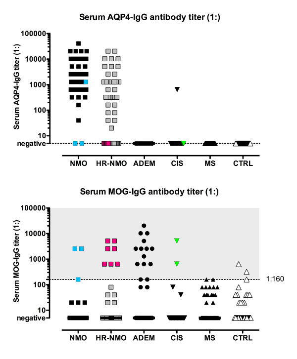
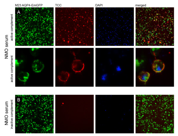
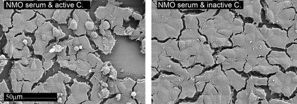
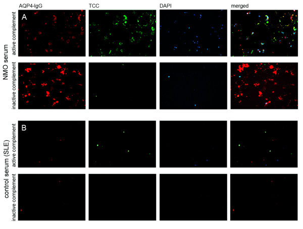
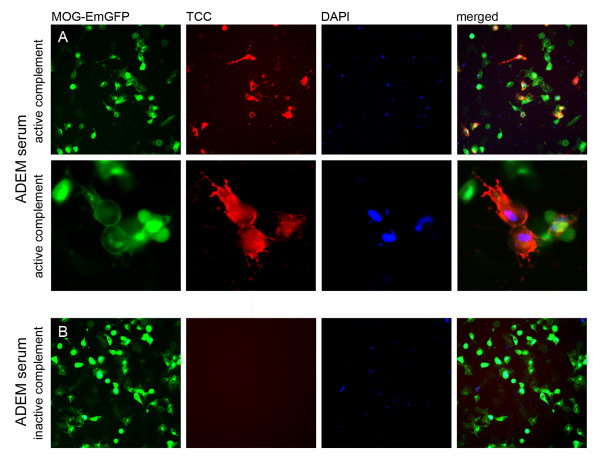
Results: We analyzed patients with definite NMO (n = 45), HR-NMO (n = 53), ADEM (n = 33), clinically isolated syndromes presenting with myelitis or optic neuritis (CIS, n = 32), MS (n = 71) and controls (n = 101; 24 other neurological diseases-OND, 27 systemic lupus erythematosus-SLE and 50 healthy subjects) for serum IgG to MOG and AQP4. Furthermore, we investigated whether these antibodies can mediate complement dependent cytotoxicity (CDC). AQP4-IgG was found in patients with NMO (n = 43, 96%), HR-NMO (n = 32, 60%) and in one CIS patient (3%), but was absent in ADEM, MS and controls. High-titer MOG-IgG was found in patients with ADEM (n = 14, 42%), NMO (n = 3, 7%), HR-NMO (n = 7, 13%, 5 rON and 2 LETM), CIS (n = 2, 6%), MS (n = 2, 3%) and controls (n = 3, 3%, two SLE and one OND). Two of the three MOG-IgG positive NMO patients and all seven MOG-IgG positive HR-NMO patients were negative for AQP4-IgG. Thus, MOG-IgG were found in both AQP4-IgG seronegative NMO patients and seven of 21 (33%) AQP4-IgG negative HR-NMO patients. Antibodies to MOG and AQP4 were predominantly of the IgG1 subtype, and were able to mediate CDC at high-titer levels.

Conclusions: We could show for the first time that a subset of AQP4-IgG seronegative patients with NMO and HR-NMO exhibit a MOG-IgG mediated immune response, whereas MOG is not a target antigen in cases with an AQP4-directed humoral immune response.


Figures

Figure 1
Figure 1
Serum MOG-IgG and AQP4-IgG antibody titer levels in CNS demyelinating diseases and controls. Serum AQP4-IgG (upper panel) were exclusively detected in serum samples of patients with NMO and HR-NMO and one CIS patient, but not in any subjects with ADEM, MS and CTRL. Serum MOG-IgG (lower panel) were present at highest titers (≥ 1:160, indicated by a dotted vertical line) in a cohort of patients with ADEM, AQP4-IgG seronegative HR-NMO (pink squares) and in both AQP4-IgG seronegative NMO subjects (blue squares, both at a MOG-IgG titer of 1:2,560) as well as in two CIS samples (green triangles). One patient (NMO) was found to be double positive for MOG-IgG (blue square at the threshold level of 1:160, lower panel) and AQP4-IgG (blue square at a titer of 1:1,280; upper panel). Coloured icons indicate individual patients in the upper and lower panel.
Figure 2
Figure 2
Longitudinally extending spinal cord lesions in a high-titer MOG-IgG seropositive and AQP4-IgG seronegative patient. Sagittal T2 weighted MRI of a lesion extending over 8 vertebral segments (upper left) in a 49 year old female patient (Table 2, patient number 10). Transversal T2 weighted image (lower left) shows the extension over the whole cross-sectional area of the spinal cord. Contrast enhanced T1 weighted images (sagittal, upper right and transversal, lower right) show scattered contrast enhancement in the lesion.
Figure 3
Figure 3
Serum AQP4-IgG antibodies of an NMO patient activate the complement cascade in the presence of active complement resulting in the deposition of the terminal C5b-9 complement complex. A. Formation of the terminal complement complex (TCC, red) on the surface of M23 AQP4-EmGFP expressing HEK-293A cells (green) following addition of a heat- inactivated AQP4-IgG positive serum sample (titer of 1:10,240). The membrane attack complex resulted in lysis of AQP4-expressing cells (DAPI, blue). B. No antibody mediated complement activation was detectable following incubation of the same heat-inactivated AQP4-IgG positive NMO serum sample supplemented with inactive complement (TCC, red). Images are representative of similar staining patterns observed in independent complement activation assays of the 27 AQP4-IgG positive patients (Table 3). Images are shown in 20 × (A-upper panel and B) and 63 × (A-lower panel) magnification.
Figure 4
Figure 4
Scanning electron microscopy of serum AQP4-IgG mediated complement activation. Following incubation of AQP4 transfected HEK-293A cells with an NMO serum (AQP4-IgG titer: 1:10,240) and either active or inactive complement, the cells were investigated via scanning electron microscopy (1,000 × magnification). NMO serum supplemented with active complement resulted in increased apoptosis (shown by the detached cells, left panel). In contrast, the cell layer of NMO serum supplemented with inactive complement remained largely preserved (right panel). Images are representative of similar observations in independent complement activation assays.
Figure 5
Figure 5
Co-localization of the terminal complement complex (TCC) with an NMO patient's serum AQP4-IgG. A. The heat-inactivated serum sample of an AQP4-IgG positive NMO patient (titer 1:5,120) was added to M23 AQP4 transfected cells (without EmGFP fusion protein) in the presence of either active or inactive complement. Complement-mediated cytotoxicity (TCC, green) was only detectable after addition of active complement. Furthermore, the TCC co-localized (merged) with the antibodies directed to AQP4 (AQP4-IgG, red) resulting in lysis of the AQP4 transfected cells (DAPI staining, blue). The activation of the complement cascade was accompanied by an internalization of AQP4-IgG, resulting in an attenuated signal (red). B. An AQP4-IgG negative serum sample of an SLE patient resulted in no formation of the TCC in the presence of active complement.
Figure 6
Figure 6
Serum MOG-IgG mediated complement activation. A. High-titer MOG-IgG antibodies of an ADEM patient (titer 1:20,480) can activate the complement system on MOG expressing HEK-293A cells (MOG-EmGFP, green), resulting in TCC formation (red) and cell lysis (blue) following addition of active complement. B. No TCC formation was observed with the MOG-IgG positive serum when incubated with inactive complement, resulting in fewer DAPI stained cells (blue). Images are representative of similar staining patterns observed in independent complement activation assays of the 17 MOG-IgG positive patients (Table 3). Images are shown in 20 × (A-upper panel and B) and 63 × (A-lower panel) magnification.

References

    1. Lennon VA, Kryzer TJ, Pittock SJ, Verkman AS, Hinson SR. IgG marker of optic-spinal multiple sclerosis binds to the aquaporin-4 water channel. J Exp Med. 2005;202(4):473–477. doi: 10.1084/jem.20050304. - DOI - PMC - PubMed
    1. Lennon VA, Wingerchuk DM, Kryzer TJ, Pittock SJ, Lucchinetti CF, Fujihara K, Nakashima I, Weinshenker BG. A serum autoantibody marker of neuromyelitis optica: distinction from multiple sclerosis. Lancet. 2004;364(9451):2106–2112. doi: 10.1016/S0140-6736(04)17551-X. - DOI - PubMed
    1. Wingerchuk DM, Lennon VA, Pittock SJ, Lucchinetti CF, Weinshenker BG. Revised diagnostic criteria for neuromyelitis optica. Neurology. 2006;66(10):1485–1489. doi: 10.1212/01.wnl.0000216139.44259.74. - DOI - PubMed
    1. Satoh J, Tabunoki H, Yamamura T, Arima K, Konno H. Human astrocytes express aquaporin-1 and aquaporin-4 in vitro and in vivo. Neuropathology. 2007;27(3):245–256. doi: 10.1111/j.1440-1789.2007.00774.x. - DOI - PubMed
    1. Neely JD, Christensen BM, Nielsen S, Agre P. Heterotetrameric composition of aquaporin-4 water channels. Biochemistry. 1999;38(34):11156–11163. doi: 10.1021/bi990941s. - DOI - PubMed

Publication types

MeSH terms