. 2011 Dec;2(12):1227-43.
doi: 10.18632/oncotarget.412.

Characterization of the human folate receptor alpha via novel antibody-based probes

Affiliations

Characterization of the human folate receptor alpha via novel antibody-based probes

Daniel J O'Shannessy et al. Oncotarget. 2011 Dec.

Abstract

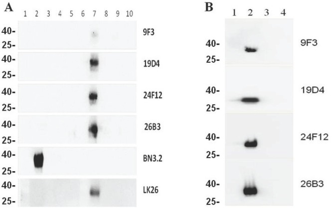
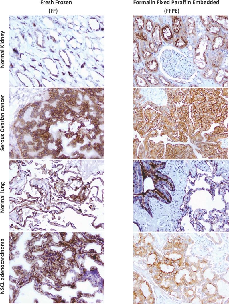
Folate receptor alpha (FRA) is a cell surface protein whose aberrant expression in malignant cells has resulted in its pursuit as a therapeutic target and marker for diagnosis of cancer. The development of immune-based reagents that can reproducibly detect FRA from patient tissue processed by varying methods has been difficult due to the complex post-translational structure of the protein whereby most reagents developed to date are highly structure-sensitive and have resulted in equivocal expression results across independent studies. The aim of the present study was to generate novel monoclonal antibodies (mAbs) using modified full length FRA protein as immunogen in order to develop a panel of mAbs to various, non-overlapping epitopes that may serve as diagnostic reagents able to robustly detect FRA-positive disease. Here we report the development of a panel of FRA-specific mAbs that are able to specifically detect FRA using an array of diagnostic platforms and methods. In addition, the methods used to develop these mAbs and their diverse binding properties provide additional information on the three dimensional structure of FRA in its native cell surface configuration.


Figures

Figure 1
Figure 1. Fluorescence Activated Cell Sorting (FACS)
Antibodies were tested for their ability to detect FRA on recombinant CHO cells expressing human FRA, FRB or FRD. Twenty of the 69 mAbs developed here were able to specifically recognize FRA and not the other folate receptor orthologs or negative controls. Shown is a representative analysis of lead antibodies and their robust FRA-specific binding. We then tested the ability of these antibodies to cross react to epitopes or with the previously reported anti-FRA mAb MORAb-003 which is being pursued in clinical development for the treatment of ovarian cancer [20]. Very few mAbs were found to compete for binding. Of particular note was the cross reactivity of 24F12 with 26B3 and 19D4 with MORAb-003 (not shown).
Figure 2
Figure 2. Western blotting of mAbs to reduced and non-reduced FR isoforms
Purified recombinant (panel A) and whole cell lysates (panel B) from CHO cells expressing FRA or FR homologs FRB, FRD or FRG were run on SDS-PAGE gels. Proteins were prepared in sample buffer with or without reducing agents. Panel A, lane 1, molecular weight markers, lanes 2-5. 0.5μg reduced FRA, FRB, FRG, and FRD, respectively; lane 6, blank; lanes 7-10, 0.5μg non-reduced FRA, FRB, FRG, and FRD, respectively. The positive band represents the only reactive species in each lane and corresponds to a molecular weight of ~38kDa. Panel B, lane 1 molecular weight markers, lane 2 CHO-FRA, lane 3, CHO-FRB, lane 4 CHO-FRD whole cell lysates prepared in sample buffer without reducing agents on a SDS-PAGE gel. Each panel is probed with the designated anti-FRA mAb labeled on the right. The molecular weights for FR are: FRA ~38kDA; FRB ~30kDa; FRG ~28kDa; FRD ~26kDa.
Figure 3
Figure 3. IHC of mAbs on human tissues
mAbs were used to assess the ability to detect FRA in tissues prepared via FFPE or FF methods. Shown is a representative analysis of anti-FRA mAbs developed here that demonstrate specific staining of FRA positive tissues prepared using various methods. mAb 26B3 showed the most robust staining for FRA in IHC and is presented above. Shown is the ability of 26B3 to detect FRA in normal tissues and previously identified FRA-positive cancers (serous ovarian cancer and NSCLC adenocarcinoma) prepared using formalin fixed paraffin embedded (FFPE, right column) and fresh frozen (FF, left column) tissue. As shown, mAb 26B3 is able to robustly recognize antigen in both tissue preparations. Slides shown at 20X magnification.
Figure 4
Figure 4. Design of H/D mapping strategy
To map regions of the free FRA protein or protein bound to mAb, the protein is incubated with “on solution” that exchanges external hydrogens (depicted by blue circles) with deuterium ions (red circles). Upon saturation, the deuterated FRA is then incubated in H2O which exchanges hydrogen for deuterium in unbound regions or regions no longer protected by subsequent mAb binding. As control, “on-column” reactions are conducted whereby non-deuterated FRA is bound by mAb and then deuterated followed by hydrogen exchange to identify protected region bound by the anti-FRA mAb. To identify external regions on the free FRA protein, reactions are conducted with FRA at varying time points. The green circles represent the FRA protein. The yellow cylinder represents the anti-FRA antibody binding region which inhibits the hydrogen or deuterium exchange on the deuterated or non-deuterated FRA protein, respectively.
Figure 5
Figure 5. Hydrogen/deuterium exchange map of the recombinant human folate receptor alpha (rFRA) in solution
H/D exchange map of rFRA at 23°C at pH7.0. Each block represents overlapping peptides analyzed. Each block contains 5 time points, as indicated on the legend to the right. Deuteration level at each time point on each peptide is indicated on the legend as well. For externally exposed regions fast H/D rates at the 30s time point that increase linearly with extended incubation periods indicate more externally exposed surfaces [32]. As shown here, amino acid regions 45-57, 129-142 and 174-184 indicate the most externally accessible regions on the FRA molecule.
Figure 6
Figure 6. H/D mapping of anti-FRA mAb epitopes via hydrogen/deuteration exchange of FRA upon antibody complexation
Shown in Panel A are the perturbations of H/D exchange of rFRA upon antibody binding, illustrated as a heat map for each antibody. The level of perturbation, as a percentage, is shown in the legend in the lower right. Included above is the H/D exchange map of free rFRA in solution for comparison and alignment of the proposed epitope regions of 24F12, 9F3, 26B3 and MORAb-003 mAbs, respectively. For MORAb-003, epitopes were confirmed via di-alanine scanning of full length rFRA vs mutant FRA variants which refined the epitope for the antibody from the 45-57 region to amino acid residues HKDV (Panel B). The H/D mapped epitope regions for each mAb is identified and provided in Table 4. Panel A. H/D exchange map of anti-FRA mAbs on rFRA at varying incubation time points. The antibody for each exchange map is shown on the left and the amino acid sequence of FRA is shown on the top. Panel B. Di-alanine scanning of FRA of the MORAb-003 epitope region 45-57 identified via H/D mapping was conducted to further refine the epitope of the antibody. MORAb-003 had reduced binding of FRAs containing changes within the HKDV amino acids as determined via surface plasmon resonance. Shown are the 12 mutant di-alanine full length FRA mutants and the binding characteristics of MORAb-003 to each mutant with values shown on the right.
Figure 7
Figure 7. Modeling of the anti-FRA antibody epitopes on the FRA-related structure, chicken riboflavin-binding protein (cRBP)
Sequences of cRBP and hFRA were aligned and linear sequences of anti-FRA antibody epitopes as well as external domains of the free FRA protein were mapped on cRBP and highlighted on the cRBP 3-D structure. The model is shown from two different angles to depict the epitopes for each antibody as well as the free protein analyzed by H/D mapping. As anticipated by the H/D method, those regions showing highest H/D exchange are localized to the external regions of the FRA protein.

References

    1. Choi SW, Mason JB. Folate and carcinogenesis: an integrated scheme. J Nutr. 2000;130:129–32. - PubMed
    1. Matherly LH, Goldman DI. Membrane transport of folates. Vitam Horm. 2003;66:403–56. - PubMed
    1. Elnakat H, Ratnam M. Distribution, functionality and gene regulation of folate receptor isoforms: implications in targeted therapy. Adv Drug Deliv Rev. 2004;56:1067–84. - PubMed
    1. Basal E, Eghbali-Fatourechi GZ, Kalli KR, Hartmann LC, Goodman KM, Goode EL, Kamen BA, Low PS, Knutson KL. Functional folate receptor alpha is elevated in the blood of ovarian cancer patients. PLoS One. 2009;4:e6292. - PMC - PubMed
    1. Reddy JA, Haneline LS, Srour EF, Antony AC, Clapp DW, Low PS. Expression and functional characterization of the beta-isoform of the folate receptor on CD34(+) cells. Blood. 1999;93:3940–48. - PubMed

MeSH terms