Comparative Study
. 2012 Feb 21;53(2):873-80.
doi: 10.1167/iovs.11-8611. Print 2012 Feb.

Depth-dependent transverse shear properties of the human corneal stroma

Affiliations
Comparative Study

Depth-dependent transverse shear properties of the human corneal stroma

Steven J Petsche et al. Invest Ophthalmol Vis Sci. .

Abstract

Purpose: To measure the transverse shear modulus of the human corneal stroma and its profile through the depth by mechanical testing, and to assess the validity of the hypothesis that the shear modulus will be greater in the anterior third due to increased interweaving of lamellae.

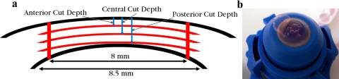
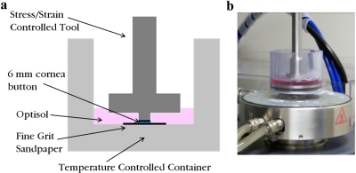
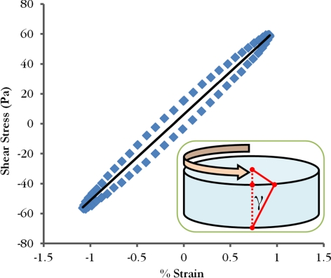
Methods: Torsional rheometry was used to measure the transverse shear properties of 6 mm diameter buttons of matched human cadaver cornea pairs. One cornea from each pair was cut into thirds through the thickness with a femtosecond laser and each stromal third was tested individually. The remaining intact corneas were tested to measure full stroma shear modulus. The shear modulus from a 1% shear strain oscillatory test was measured at various levels of axial compression for all samples.

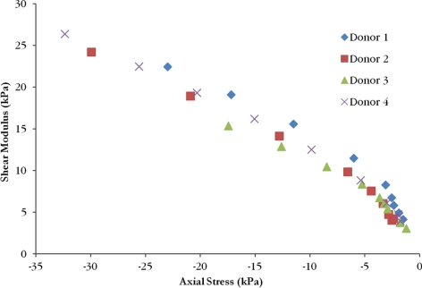
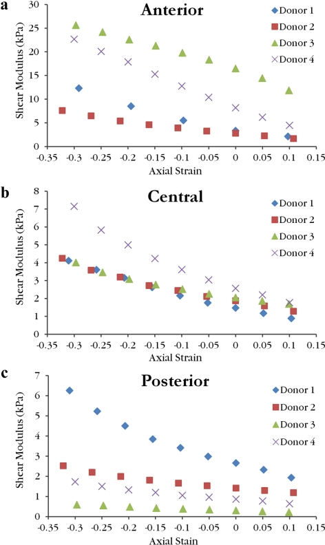
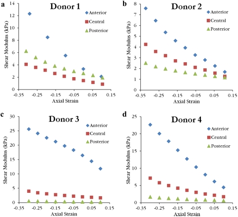
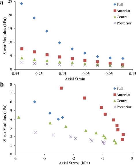
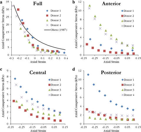
Results: After controlling for axial compression, the transverse shear moduli of isolated anterior layers were significantly higher than central and posterior layers. Mean modulus values at 0% axial strain were 7.71 ± 6.34 kPa in the anterior, 1.99 ± 0.45 kPa in the center, 1.31 ± 1.01 kPa in the posterior, and 9.48 ± 2.92 kPa for full thickness samples. A mean equilibrium compressive modulus of 38.7 ± 8.6 kPa at 0% axial strain was calculated from axial compression measured during the shear tests.

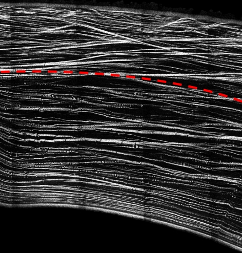
Conclusions: Transverse shear moduli are two to three orders of magnitude lower than tensile moduli reported in the literature. The profile of shear moduli through the depth displayed a significant increase from posterior to anterior. This gradient supports the hypothesis and corresponds to the gradient of interwoven lamellae seen in imaging of stromal cross-sections.


Figures

Figure 1.
Figure 1.
Cross-section of a human cornea from second harmonic generated imaging showing the interweaving of lamellae in the anterior third.
Figure 2.
Figure 2.
Four cuts were made with the femtosecond laser, three 8-mm diameter raster cuts at different depths and one 8.5-mm diameter circular cut through the thickness (a). An anterior third is shown in the process of separation immediately before testing (b).
Figure 3.
Figure 3.
A schematic of the experimental setup indicates the position of the upper tool when the corneal button is tested in the torsional rheometer (a). The corneal storage medium (Optisol; Bausch & Lomb) bath was added after the upper tool had been lowered to the desired gap height for the first test. The photograph of the experimental setup shows the heating plate below the sample that keeps the bath at 37°C (b).
Figure 4.
Figure 4.
Typical stress/strain curve from a final load cycle used for calculating the magnitude of the complex shear modulus. The inset cartoon shows the applied torque and resulting shear strain (γ) on a cornea button, which is maximum at the perimeter.
Figure 5.
Figure 5.
The shear moduli as a function of axial compressive stress for the full thickness samples from each donor displays a nonlinear relationship.
Figure 6.
Figure 6.
The shear moduli as a function of axial strain for the anterior (a), central (b), and posterior (c) isolated layer samples reveal the variation of moduli range between layers. Note the scale of the ordinate axis is larger for anterior samples when compared with central and posterior samples.
Figure 7.
Figure 7.
The shear moduli as a function of axial strain for the laser cut corneas from Donor 1 (a), Donor 2 (b), Donor 3 (c), and Donor 4 (d) illustrate that the anterior layer was stiffer than the central and posterior layers at all compressive strains. Also, except for Donor 1, the central layer was stiffer than the posterior layer at every compressive strain.
Figure 8.
Figure 8.
All shear moduli from Donor 2 are plotted as a function of axial strain (a) and axial compressive stress (b). The full thickness sample is stiffer than the isolated layer samples for a given axial strain. Full sample modulus values fall between the anterior and central samples at a given axial stress. On both graphs, moving left corresponds to increasing compression.
Figure 9.
Figure 9.
The equilibrium axial compressive stress (swelling pressure) as a function of axial strain follows a power law relationship. A power fit reported by Olsen and Sperling is transformed to axial strain for comparison to full thickness samples (a). Data are believed to lie below this curve as a result of the corneal storage medium (Optisol; Bausch & Lomb) bath. Swelling pressure values for anterior (b), central (c), and posterior (d) isolated layers are low compared with the full thickness sample.

Similar articles

Cited by

References

    1. Elsheikh A, Wang D, Brown M, Rama P, Campanelli M, Pye D. Assessment of corneal biomechanical properties and their variation with age. Curr Eye Res. 2007;32:11–19 - PubMed
    1. Elsheikh A, Brown M, Alhasso D, Rama P, Campanelli M, Garway-Heath D. Experimental assessment of corneal anisotropy. J Refract Surg. 2008;24:178–187 - PubMed
    1. Hjortdal JØ. Regional elastic performance of the human cornea. J Biomech. 1996;29:931–942 - PubMed
    1. Boyce BL, Grazier JM, Jones RE, Nguyen TD. Full-field deformation of bovine cornea under constrained inflation conditions. Biomaterials. 2008;29:3896–3904 - PubMed
    1. Christensen RM. Chapter 3.1: Transversely isotropic media. In: Mechanics of Composite Materials. 1st ed New York, NY: John Wiley & Sons; 1979:74–80

Publication types