

Effects of insulin-like growth factor-1 on the properties of mesenchymal stem cells in vitro
- PMID: 22205616
- PMCID: PMC3251748
- DOI: 10.1631/jzus.B1100117
Effects of insulin-like growth factor-1 on the properties of mesenchymal stem cells in vitro
Abstract
Objective: To explore the effects of insulin-like growth factor-1 (IGF-1) on migration, proliferation and differentiation of mesenchymal stem cells (MSCs).
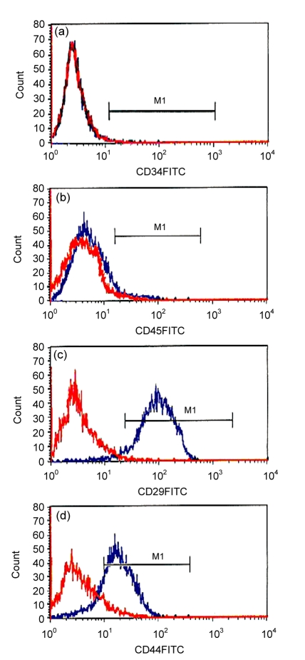
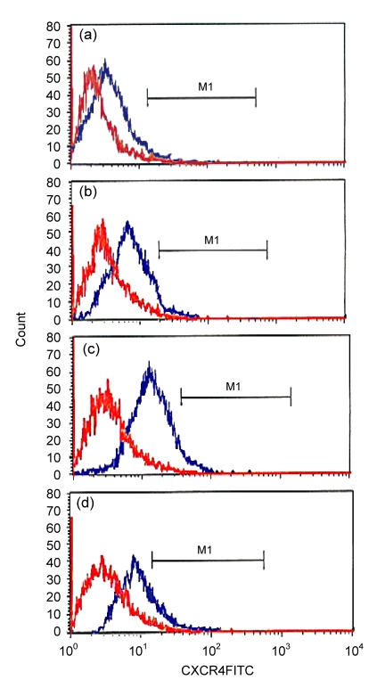
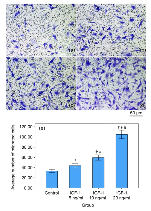
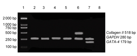
Methods: MSCs were obtained from Sprague-Dawley rats by a combination of gradient centrifugation and cell culture techniques and treated with IGF-1 at concentrations of 5-20 ng/ml. Proliferation of MSCs was determined as the mean doubling time. Expression of CXC chemokine receptor 4 (CXCR4) and migration property were determined by flow cytometry and transwell migration essay, respectively. mRNA expression of GATA-4 and collagen II was determined by reverse transcription-polymerase chain reaction (RT-PCR).
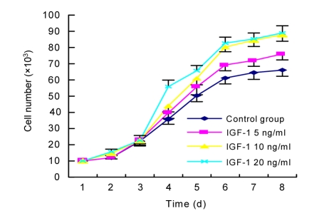
Results: The mean doubling time of MSC proliferation was decreased, and the expression of CXCR4 on MSCs and migration of MSCs were increased by IGF-1, all in a dose-dependent manner, while the optimal concentration of IGF-1 on proliferation and migration was different. IGF-1 did not affect the expression of GATA-4 or collagen II mRNA.
Conclusions: IGF-1 dose-dependently stimulated the proliferation of MSCs, upregulated the expression of CXCR4, and accelerated migration. There was no apparent differentiation of MSCs to cardiomyocytes or chondrocytes after culturing with IGF-1 alone.
Figures
Similar articles
-
Tanshinone IIA and astragaloside IV promote the migration of mesenchymal stem cells by up-regulation of CXCR4.Protoplasma. 2013 Apr;250(2):521-30. doi: 10.1007/s00709-012-0435-1. Epub 2012 Aug 8. Protoplasma. 2013. PMID: 22872094
-
Effect of IGF-I in the chondrogenesis of bone marrow mesenchymal stem cells in the presence or absence of TGF-beta signaling.J Bone Miner Res. 2006 Apr;21(4):626-36. doi: 10.1359/jbmr.051213. Epub 2006 Apr 5. J Bone Miner Res. 2006. PMID: 16598383
-
Hepatocyte growth factor combined with insulin like growth factor-1 improves expression of GATA-4 in mesenchymal stem cells cocultured with cardiomyocytes.Chin Med J (Engl). 2008 Feb 20;121(4):336-40. Chin Med J (Engl). 2008. PMID: 18304467
-
Study on the mechanism of quercetin inducing mesenchymal stem cells to differentiate into fibroblasts through TGF-β1 and IGF-1.Tissue Cell. 2024 Jun;88:102383. doi: 10.1016/j.tice.2024.102383. Epub 2024 Apr 11. Tissue Cell. 2024. PMID: 38613933
-
The surface adhesion molecule CXCR4 stimulates mesenchymal stem cell migration to stromal cell-derived factor-1 in vitro but does not decrease apoptosis under serum deprivation.Cardiovasc Revasc Med. 2006 Jan-Mar;7(1):19-24. doi: 10.1016/j.carrev.2005.10.008. Cardiovasc Revasc Med. 2006. PMID: 16513519
Cited by
-
The effect of Kisspeptin-10 on mesenchymal stem cells migration in vitro and in vivo.Adv Biomed Res. 2015 Jan 30;4:20. doi: 10.4103/2277-9175.149851. eCollection 2015. Adv Biomed Res. 2015. PMID: 25709985 Free PMC article.
-
Optimization of Media Change Intervals through Hydrogels Using Mathematical Models.Biomacromolecules. 2023 Feb 13;24(2):604-612. doi: 10.1021/acs.biomac.2c00961. Epub 2023 Feb 1. Biomacromolecules. 2023. PMID: 36724373 Free PMC article.
-
G9a inhibition promotes the formation of pacemaker-like cells by reducing the enrichment of H3K9me2 in the HCN4 promoter region.Mol Med Rep. 2023 Feb;27(2):21. doi: 10.3892/mmr.2022.12908. Epub 2022 Dec 9. Mol Med Rep. 2023. PMID: 36484369 Free PMC article.
-
The effects of kisspeptin-10 on migration and proliferation of endothelial cell.Adv Biomed Res. 2015 Feb 11;4:41. doi: 10.4103/2277-9175.151250. eCollection 2015. Adv Biomed Res. 2015. PMID: 25789267 Free PMC article.
-
Influence of Dipeptidyl Peptidase-4 (DPP4) on Mesenchymal Stem-Cell (MSC) Biology: Implications for Regenerative Medicine - Review.Stem Cell Rev Rep. 2022 Jan;18(1):56-76. doi: 10.1007/s12015-021-10285-w. Epub 2021 Oct 22. Stem Cell Rev Rep. 2022. PMID: 34677817 Review.
References
-
- Bartunek J, Croissant JD, Wijns W, Gofflot S, de Lavareille A, Vanderheyden M, Kaluzhny Y, Mazouz N, Willemsen P, Penicka M, et al. Pretreatment of adult bone marrow mesenchymal stem cells with cardiomyogenic growth factors and repair of the chronically infarcted myocardium. Am J Physiol Heart Circ Physiol. 2007;292(2):H1095–H1104. doi: 10.1152/ajpheart.01009.2005. - DOI - PubMed
-
- Chen SL, Fang WW, Ye F, Liu YH, Qian J, Shan SJ, Zhang JJ, Chunhua RZ, Liao LM, Lin S, et al. Effect on left ventricular function of intracoronary transplantation of autologous bone marrow mesenchymal stem cell in patients with acute myocardial infarction. Am J Cardiol. 2004;94(1):92–95. doi: 10.1016/j.amjcard.2004.03.034. - DOI - PubMed
-
- Kaplan RC, Strickler HD, Rohan TE, Muzumdar R, Brown DL. Insulin-like growth factors and coronary heart disease. Cardiol Rev. 2005;13(1):35–39. - PubMed
-
- Katritsis DG, Sotiropoulou PA, Karvouni E, Karabinos I, Korovesis S, Perez SA, Voridis EM, Papamichail M. Transcoronary transplantation of autologous mesenchymal stem cells and endothelial progenitors into infarcted human myocardium. Catheter Cardiovasc Interv. 2005;65(3):321–329. doi: 10.1002/ccd.20406. - DOI - PubMed
Publication types
MeSH terms
Substances
LinkOut - more resources
Full Text Sources
Other Literature Sources
Miscellaneous
