. 2012 Mar;86(5):2817-25.
doi: 10.1128/JVI.05685-11. Epub 2011 Dec 28.

Influenza A virus infection results in a robust, antigen-responsive, and widely disseminated Foxp3+ regulatory T cell response

Affiliations

Influenza A virus infection results in a robust, antigen-responsive, and widely disseminated Foxp3+ regulatory T cell response

Richard J Betts et al. J Virol. 2012 Mar.

Abstract

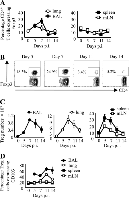
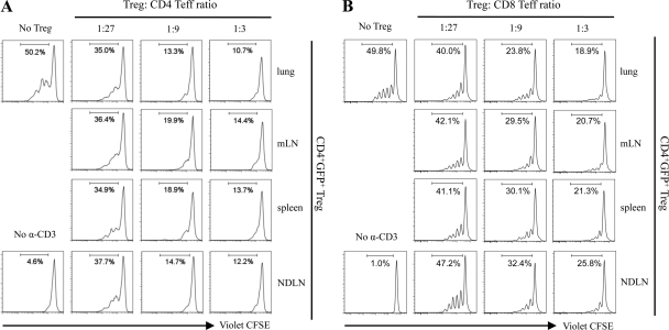
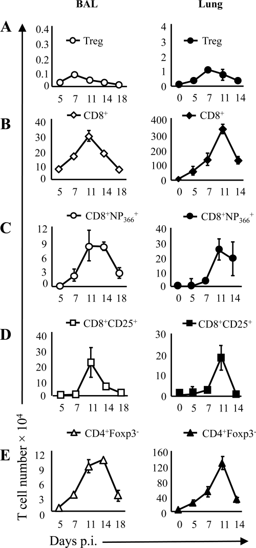
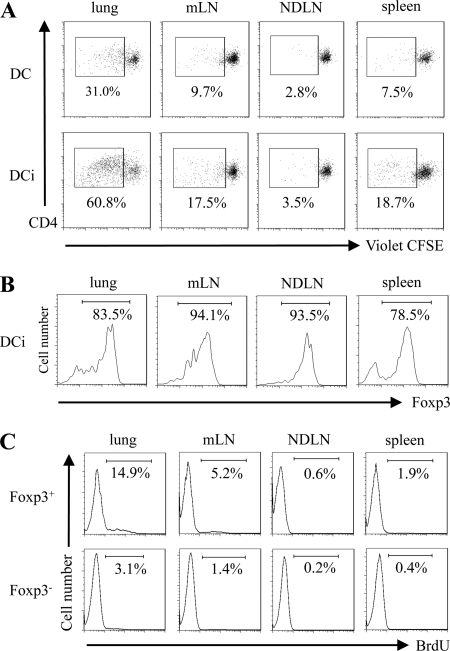
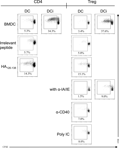
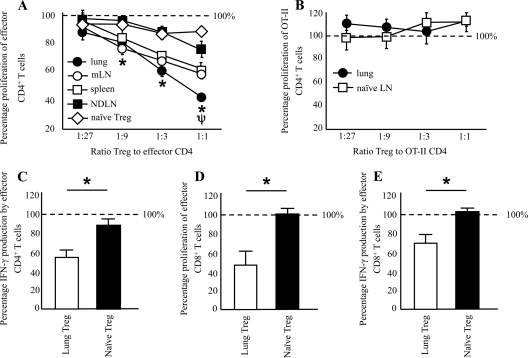
Foxp3(+) CD4(+) regulatory T cells (Tregs) represent a highly suppressive T cell subset with well-characterized immunosuppressive effects during immune homeostasis and chronic infections, although the role of these cells in acute viral infections is poorly understood. The present study sought to examine the induction of Foxp3(+) CD4(+) Tregs in a nonlethal murine model of pulmonary viral infection by the use of the prototypical respiratory virus influenza A. We establish that influenza A virus infection results in a robust Foxp3(+) CD4(+) T cell response and that regulatory T cell induction at the site of inflammation precedes the effector T cell response. Induced Foxp3(+) CD4(+) T cells are highly suppressive ex vivo, demonstrating that influenza virus-induced Foxp3(+) CD4(+) T cells are phenotypically regulatory. Influenza A virus-induced regulatory T cells proliferate vigorously in response to influenza virus antigen, are disseminated throughout the site of infection and primary and secondary lymphoid organs, and retain Foxp3 expression in vitro, suggesting that acute viral infection is capable of inducing a foreign-antigen-specific Treg response. The ability of influenza virus-induced regulatory T cells to suppress antigen-specific CD4(+) and CD8(+) T cell proliferation and cytokine production correlates closely to their ability to respond to influenza virus antigens, suggesting that virus-induced Tregs are capable of attenuating effector responses in an antigen-dependent manner. Collectively, these data demonstrate that primary acute viral infection is capable of inducing a robust, antigen-responsive, and suppressive regulatory T cell response.


Figures

Fig 1
Fig 1
Influenza A virus infection results in a robust Foxp3+ CD4+ T cell response. Mice were infected with a sublethal dose of influenza virus A/PR/8/34, and Foxp3+ CD4+ T cell proportions and numbers in the BAL fluid, lung parenchyma, mediastinal lymph node, and spleen were examined. (A) Percentage of CD3+ CD4+ cells expressing Foxp3+ in BAL fluid, lung, spleen, and mLN across a time course following influenza virus infection. (B) Representative fluorescence-activated cell sorter (FACS) plots of the proportion of CD4+ cells expressing Foxp3 in the BAL compartment, gated on lymphocyte population and CD3+ cells. Numbers represent percentages of CD4+ T cells coexpressing Foxp3. (C) Total Foxp3+ CD3+ CD4+ T cell numbers across a time course following influenza A virus infection. (D) Percentage of Foxp3+ CD3+ CD4+ T cells coexpressing Treg activation marker CD103 in BAL fluid, lung parenchyma, mLN, and spleen across a time course following influenza A virus infection. n = 3 to 9 for all; data represent means ± standard errors of the means (SEM).
Fig 2
Fig 2
Foxp3+ CD4+ T cells induced during influenza A virus infection are highly suppressive. (A) Foxp3+ CD4+ T cells were sorted from whole lung, mLN, spleen, and NDLN on the basis of eGFP expression from the lungs of influenza virus-infected mice at day 8 postinoculation and cultured in increasing proportions with violet CFSE-labeled Foxp3 CD4+ responder cells collected from naïve mice and irradiated splenocytes for 3 days, after which responder cell proliferation was examined. (B) Naïve CD8+ T cells were used as responder T cells. The number in each plot represents the percentage of cells that had gone through at least one cell cycle division. Results are representative of 3 independent experiments for panel A and 2 independent experiments for panel B.
Fig 3
Fig 3
The peak Foxp3+ CD4+ T cell response occurs prior to maximal adaptive immune response within the lung. Foxp3+ CD4+ T cell numbers in BAL fluid and lung parenchyma (A) correlated with total CD8+ T cell (B), NP366+ CD8+ T cell (C), CD25+ CD8+ T cell (D), and Foxp3 CD4+ T cell (E) numbers across a time course following influenza A virus infection. n = 3 to 6 for all; data represent means ± SEM.
Fig 4
Fig 4
Influenza A virus infection-induced Foxp3+ CD4+ T cells are antigen responsive and widely disseminated and stably express Foxp3. (A) Foxp3+ CD4+ T cells were collected from whole lung, mLN, spleen, and NDLN of influenza A virus-infected mice at day 8 postinoculation, stained with violet CFSE, and cultured with BMDCs in the presence (DCi) or absence (DC) of influenza A virus for 4 days, after which proliferation was examined by FACS analysis. (B) Stability of Foxp3 expression in proliferating Treg collected from whole lung, mLN, NDLN, and spleen of influenza virus-infected mice following ex vivo stimulation with influenza virus-infected BMDCs. (C) Influenza A virus-infected mice at day 8 postinoculation were administered 3 mg of BrdU 6 h prior to euthanasia, after which the proportion of Foxp3+ CD4+ and Foxp3 CD4+ T cells incorporating BrdU from whole lung, mLN, spleen, and NDLN was determined. Data are representative of the results of 2 to 4 independent studies.
Fig 5
Fig 5
Influenza virus-induced lung Tregs proliferate in response to an influenza virus MHC class II epitope. Foxp3+ CD4+ T cells and positive-control Foxp3 CD4+ T cells were collected from whole lung of influenza A virus-infected mice at day 11 postinoculation, stained with violet CFSE, and cultured with BMDCs for 4 days with (DCi) or without (DC) influenza A virus, negative-control influenza virus MHC class I influenza A epitope NP366, or MHC class II epitope HA126–138 in the presence or absence of IA/IE MHC class II blocking antibody, agonistic α-CD40 antibody, or poly(I·C). Proliferation was examined by FACS analysis after 4 days in culture. Data are representative of the results of 3 to 4 separate experiments.
Fig 6
Fig 6
Foxp3+ CD4+ T cells induced during influenza A virus infection suppress antigen-specific CD4+ and CD8+ T cell proliferation. Foxp3+ CD4+ T cells were sorted from whole lung, mLN, and NDLN of influenza virus-infected mice at day 8 postinoculation and cultured in increasing proportions with violet CFSE-labeled Foxp3 CD4+ responder cells collected from the lungs of influenza virus-infected animals in the presence of BMDCs and influenza A virus. The number in each plot represents the percentage of cells that had gone through at least one cell cycle division. (A) Representative FACS dot plots. Plots were gated on a live T cell FS/SS population. *, P < 0.05 for each lung/mLN/spleen data point compared to the corresponding NDLN data point; ψ, P < 0.05 for each lung data point compared to the corresponding mLN/spleen data point (2-way ANOVA). (B) Cumulative data are representative of the results determined with 5 samples over 2 independent experiments with multiple mice per sample. Data represent means ± SEM. (C) Foxp3+ CD4+ T cells collected from the lungs of influenza virus-infected animals or naïve Foxp3+ CD4+ T cells were cultured in increasing proportions with violet CFSE-labeled CD4+ CD25 responder cells collected from the lymph nodes of DO11.10 mice in the presence of OVA323-339 peptide-pulsed BMDCs. Cumulative data are representative of the results determined with 5 samples over 2 independent experiments with multiple mice per sample. (D) Virus-induced CD4+ T cells from influenza virus-infected mice at day 8 postinoculation were cultured with Tregs from the lung of influenza virus-infected mice or the spleens of naïve mice for 4 days and IFN-γ levels determined by ELISA. (E) Virus-induced CD8+ T cells from influenza virus-infected mice at day 8 postinoculation were cultured with Tregs from the lung of influenza virus-infected mice or the spleens of naïve mice for 4 days, and the degree of proliferation compared to Treg-free cultures was determined by CFSE dilution. (D) IFN-γ production by virus-induced CD8+ T cells from influenza virus-infected mice at day 8 postinoculation cultured with influenza virus-induced lung Tregs or naïve compared to Treg-free cultures. (D and E) Data represent means ± SEM (n = 3 to 4). * denotes P < 0.01 (Student's t test).

References

    1. Accapezzato D, et al. 2004. Hepatic expansion of a virus-specific regulatory CD8(+) T cell population in chronic hepatitis C virus infection. J. Clin. Invest. 113:963–972 - PMC - PubMed
    1. Antunes I, et al. 2008. Retrovirus-specificity of regulatory T cells is neither present nor required in preventing retrovirus-induced bone marrow immune pathology. Immunity 29:782–794 - PMC - PubMed
    1. Belkaid Y. 2008. Role of Foxp3-positive regulatory T cells during infection. Eur. J. Immunol. 38:918–921 - PMC - PubMed
    1. Belkaid Y, Piccirillo CA, Mendez S, Shevach EM, Sacks DL. 2002. CD4+CD25+ regulatory T cells control Leishmania major persistence and immunity. Nature 420:502–507 - PubMed
    1. Belz GT, Stevenson PG, Doherty PC. 2000. Contemporary analysis of MHC-related immunodominance hierarchies in the CD8+ T cell response to influenza A viruses. J. Immunol. 165:2404–2409 - PubMed

Publication types