. 2012 Mar;86(6):3318-26.
doi: 10.1128/JVI.06566-11. Epub 2011 Dec 28.

Self-assembly of viral capsid protein and RNA molecules of different sizes: requirement for a specific high protein/RNA mass ratio

Affiliations

Self-assembly of viral capsid protein and RNA molecules of different sizes: requirement for a specific high protein/RNA mass ratio

Ruben D Cadena-Nava et al. J Virol. 2012 Mar.

Erratum in

Abstract

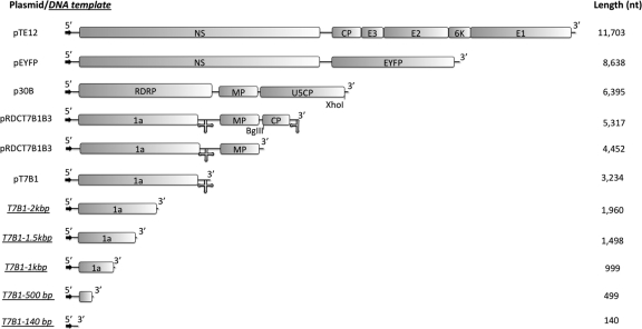
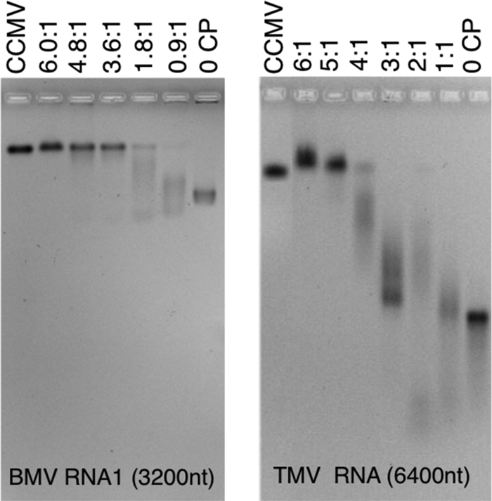
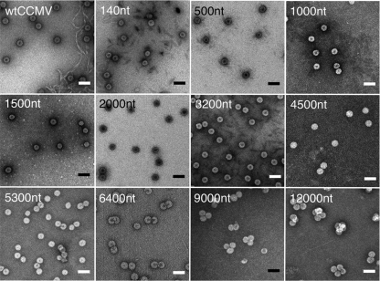
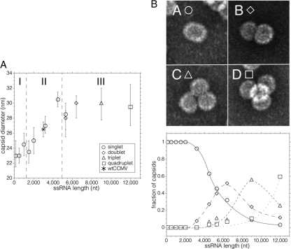
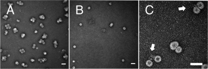
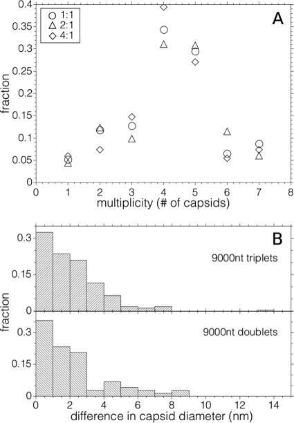
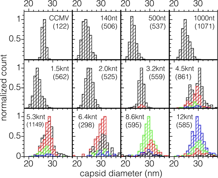
Virus-like particles can be formed by self-assembly of capsid protein (CP) with RNA molecules of increasing length. If the protein "insisted" on a single radius of curvature, the capsids would be identical in size, independent of RNA length. However, there would be a limit to length of the RNA, and one would not expect RNA much shorter than native viral RNA to be packaged unless multiple copies were packaged. On the other hand, if the protein did not favor predetermined capsid size, one would expect the capsid diameter to increase with increase in RNA length. Here we examine the self-assembly of CP from cowpea chlorotic mottle virus with RNA molecules ranging in length from 140 to 12,000 nucleotides (nt). Each of these RNAs is completely packaged if and only if the protein/RNA mass ratio is sufficiently high; this critical value is the same for all of the RNAs and corresponds to equal RNA and N-terminal-protein charges in the assembly mix. For RNAs much shorter in length than the 3,000 nt of the viral RNA, two or more molecules are assembled into 24- and 26-nm-diameter capsids, whereas for much longer RNAs (>4,500 nt), a single RNA molecule is shared/packaged by two or more capsids with diameters as large as 30 nm. For intermediate lengths, a single RNA is assembled into 26-nm-diameter capsids, the size associated with T=3 wild-type virus. The significance of these assembly results is discussed in relation to likely factors that maintain T=3 symmetry in vivo.


Figures

Fig 1
Fig 1
Schematic representation of the plasmids and DNA templates used for RNA syntheses. The arrows denote the transcription promoter, and the cloverleaf at the 3′ end represents the highly conserved tRNA-like structure (3′TLS) in the corresponding RNA transcripts. The boxes represent the open reading frames of the RNAs; 1a is a viral replicase; MP and CP are the movement protein and capsid protein, respectively; E1, E2, E3, and 6K are structural proteins; and NS represents all nonstructural viral proteins for Sindbis virus. EYFP is the sequence that encodes for the enhanced yellow fluorescent protein, RDRP denotes the RNA-dependent RNA polymerase genes, and U5CP is the CP gene of strain U5 of tobacco mild green mosaic virus. The representations of the plasmid templates are scaled to their relative lengths.
Fig 2
Fig 2
CP-RNA assembly titrations: gel retardation assays. Shown are 1% agarose gels stained with ethidium bromide. At the left is a titration of 3,217-nt BMV RNA1 with various amounts of CCMV CP ranging from 0 (right-most lane, RNA) to the “magic” ratio, 6:1 (lane second from left). CP/RNA ratios are wt/wt ratios of CP to RNA. This gel was run in electrophoresis buffer. On the right is a similar gel retardation assay carried out with 6,395-nt TMV RNA and run in virus buffer. In both gels, the leftmost lane shows the position of wt CCMV. Note, in each gel, the appearance of a smear of intensity accompanying every assembly carried out below the magic ratio.
Fig 3
Fig 3
VLP products as a function of RNA length. Negative-stain transmission electron microscopy images of the assembly products of various lengths of ssRNA mixed with CCMV CP at the magic ratio (6:1 [wt/wt]) in RNA assembly buffer. The length of the RNA in each assembly is labeled in the upper left corner of each image. The upper leftmost image shows wt CCMV capsids. Grids were stained with uranyl acetate. Scale bars, 50 nm.
Fig 4
Fig 4
(A) Average capsid diameter as a function of RNA length. A plot of the average capsid diameter as a function of the length of ssRNA packaged, with each assembly carried out at the magic ratio in RNA assembly buffer (RAB), is shown. Each average contains contributions only from the capsids associated with the predominant multiplet (i.e., singlet, doublet, triplet, or quadruplet) for the corresponding RNA length (except in the case of 5,300 nt, where equal amounts of singlets and doublets were observed). The plot is divided along the abscissa into three regions: (I) the region between 140 and ∼1,000 nt, where we find predominantly singlet capsids that contain multiple ssRNA molecules; (II) the region between ∼1,500 and ∼4,000 nt, where each singlet capsid contains only one RNA molecule (the point denoted by an asterisk corresponds to the wt CCMV virion); and (III) the region above ∼4,000 nt corresponding to the appearance of multiplet capsids sharing an RNA molecule. The error bars represent the full width at half-maximum for each distribution. (B) Fraction of multiplets as a function of RNA length. The negative-stain TEM images at the top of the panel show typical structures observed for singlets (A), doublets (B), triplets (C), and quadruplets (D). The frequency of multiplet capsids (bottom panel)—defined as the fraction of capsids present in the transmission electron microscopy as doublets (♢), triplets (△), and quadruplets (□)—increases with ssRNA length after 3,200 nt. Singlets (○) predominate for smaller lengths. Hand-drawn best-fit lines have been added to aid the eye in following the appearance and disappearance of the different multiplet populations as RNA length increases.
Fig 5
Fig 5
Exposure of RNA in Multiplets. (A) Typical transmission electron microscopy image of 12,000-nt RNA assembled with CCMV CP. (B) Same assembly mixture after exposure to RNase A in virus buffer for 1 h. Note the reduction in the number of multiplets, as well as an overall reduction in the total number of capsids present. (C) Shared RNA between capsids in a multiplet. The arrows point to RNA seen linking two capsids. Scale bars, 50 nm.
Fig 6
Fig 6
Properties of multiplets. (A) Absence of effect of 2- and 4-fold dilutions on the frequency of multiplets. The different symbols show the effect of sample dilution on fractions of multiplets of each type. The sample represented by the circles has been diluted by factors of 2 (△) and factor of 4 (◊). There is no systematic effect on the populations of multiplets with dilution. (B) Size differences between capsids within doublets and triplets formed from the assembly of 9,000-nt RNA and CCMV capsid protein.
Fig 7
Fig 7
Distribution of capsid diameters appearing in singlets and multiplets for each length of RNA packaged. Contributions from singlets, doublets, triplets, and quadruplets are shown in black, red, green, and blue, respectively. The distribution at the upper left is for wt CCMV. In each distribution the counts have been normalized to that of the most abundant size. The total numbers of particles measured are given in parentheses.

References

    1. Adolph KW, Butler PJG. 1977. Studies on the assembly of a spherical plant virus. III. Reassembly of infectious virions under mild conditions. J. Mol. Biol. 109:345–357 - PubMed
    1. Allison RF, Janda M, Ahlquist P. 1988. Infectious in vitro transcripts from cowpea chlorotic mottle virus cDNA clones and exchange of individual RNA components with brome mosaic virus. 1988 J. Virol. 62:3581–3588 - PMC - PubMed
    1. Annamalai P, Rao ALN. 2005. Dispensability of 3′ tRNA-like sequence for packaging cowpea chlorotic mottle virus genomic RNAs. Virology 332:650–658 - PubMed
    1. Annamalai P, Rao ALN. 2006. Packaging of brome mosaic virus subgenomic RNA is functionally coupled to replication-dependent transcription and translation of coat protein. J. Virol. 80:10096–10108 - PMC - PubMed
    1. Annamalai P, Rofail F, DeMason DA, Rao ALN. 2008. Replication-coupled packaging mechanism in positive-strand RNA viruses: synchronized coexpression of functional multigenome RNA components of an animal and a plant virus in Nicotiana benthamiana cells by agroinfiltration. J. Virol. 82:1484–1490 - PMC - PubMed

Publication types