. 2012 Mar;86(5):2696-705.
doi: 10.1128/JVI.06591-11. Epub 2011 Dec 28.

Characterization of the R263K mutation in HIV-1 integrase that confers low-level resistance to the second-generation integrase strand transfer inhibitor dolutegravir

Affiliations

Characterization of the R263K mutation in HIV-1 integrase that confers low-level resistance to the second-generation integrase strand transfer inhibitor dolutegravir

Peter K Quashie et al. J Virol. 2012 Mar.

Abstract

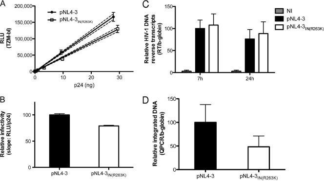
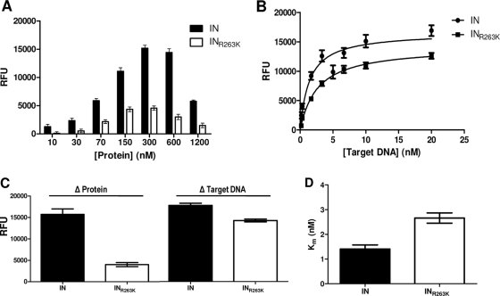
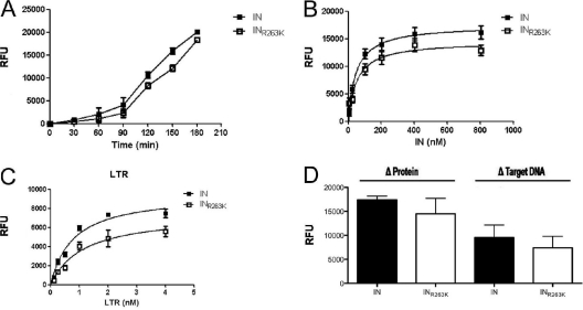
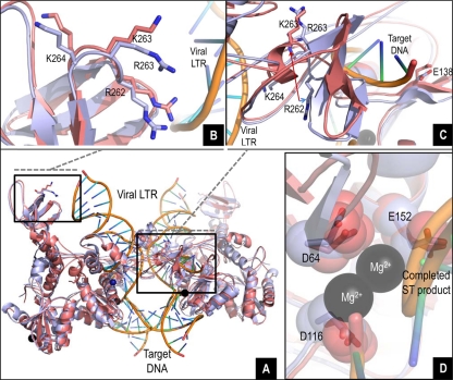
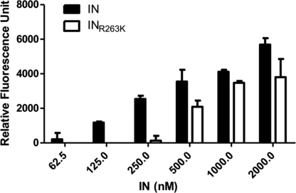
Integrase (IN) strand transfer inhibitors (INSTIs) have been developed to inhibit the ability of HIV-1 integrase to irreversibly link the reverse-transcribed viral DNA to the host genome. INSTIs have proven their high efficiency in inhibiting viral replication in vitro and in patients. However, first-generation INSTIs have only a modest genetic barrier to resistance, allowing the virus to escape these powerful drugs through several resistance pathways. Second-generation INSTIs, such as dolutegravir (DTG, S/GSK1349572), have been reported to have a higher resistance barrier, and no novel drug resistance mutation has yet been described for this drug. Therefore, we performed in vitro selection experiments with DTG using viruses of subtypes B, C, and A/G and showed that the most common mutation to emerge was R263K. Further analysis by site-directed mutagenesis showed that R263K does confer low-level resistance to DTG and decreased integration in cell culture without altering reverse transcription. Biochemical cell-free assays performed with purified IN enzyme containing R263K confirmed the absence of major resistance against DTG and showed a slight decrease in 3' processing and strand transfer activities compared to the wild type. Structural modeling suggested and in vitro IN-DNA binding assays show that the R263K mutation affects IN-DNA interactions.


Figures

Fig 1
Fig 1
The R263K mutation specifically decreases HIV-1 integration. (A and B) The R263K mutation in the IN region decreases HIV-1 pNL4-3 infectivity. (A) Relative luciferase units (RLU) produced by TZM-bl cells 48 h after infection with wild-type and IN(R263K) pNL4-3 viruses. Viral stocks were quantified by p24 ELISA and diluted as indicated. Infections were performed in triplicate for each viral dilution. Results for each triplicate are represented as means ± standard deviations (SD). The calculated linear regressions are shown as solid lines and the 95% confidence intervals as dotted lines. The results presented are representative of three independent experiments. (B) Infectivity of wild-type and R263K mutant viruses represented by the means ± SD of the calculated slopes for three independent TZM-bl infectivity assays (P < 0.01, t test), normalized against the wild-type slope, arbitrarily set at 100%. (C) The R263K mutation does not affect HIV-1 pNL4-3 reverse transcript production. Reverse transcription products were measured by qPCR at 7 h and 24 h after infection of PM1 cells with wild-type and R263K mutant viruses. Infections were performed in duplicate with two different viral stocks for each virus for a total of 4 independent infections for each time point and each virus. qPCRs were performed in duplicate for each sample. Results were normalized for β-globin gene expression and expressed relative to the normalized signal measured for the wild-type virus at 7 h postinfection, arbitrarily fixed at 100% for each set of infections. Results from noninfected cells (NI) are indicated. Means ± SD are shown. (D) HIV-1 pNL4-3 integration is diminished in the presence of the R263K mutation. Integrated DNA was quantified by qPCR in PM1 cells infected with wild-type and R263K pNL4-3 viruses for 72 h. Infections were performed twice in duplicate with two separate viral stocks, for a total of 8 independent infections for each virus. qPCRs were performed in duplicate for each sample. Results were normalized for β-globin gene expression and expressed relative to the signal detected for wild-type virus, arbitrarily set at 100% for each set of infections. Means ± SD are shown.
Fig 2
Fig 2
Strand transfer activity of purified recombinant wild-type (IN) and R263K (INR263K) integrases. (A) Strand transfer activity expressed in relative fluorescent units (RFU) in the presence of 3 nM target DNA and variable concentrations of purified recombinant protein. (B) Strand transfer activity (RFU) in the presence of 300 nM purified recombinant protein and variable concentrations of target DNA. (C) Calculated maximum strand transfer activities for wild-type IN and INR263K with variable protein or target DNA concentration. The maximum activities in the presence of increasing concentrations of proteins (Δ Protein) were calculated by excluding the higher concentration (1,200 nM). (D) Calculated Michaelis-Menten constant (Km) for purified IN and INR263K.
Fig 3
Fig 3
3′-processing activity of purified recombinant wild-type (IN) and R263K (INR263K) integrases. (A) Time-dependent 3′-processing activity expressed in relative units of time-resolved fluorescence (RFU) in the presence of 100 nM protein and 10 nM viral LTR DNA mimic (P < 0.02, paired t test). (B) 3′-processing activity (RFU) after 2 h of incubation with 10 nM target DNA and increasing concentrations of protein. (C) 3′-processing activity (RFU) after 2 h of incubation with 400 nM protein and increasing concentrations of target DNA (LTR). (D) Calculated maximum 3′-processing activities for wild-type IN and INR263K with variable protein or target DNA concentrations.
Fig 4
Fig 4
In silico studies of the wild-type and R263K integrases. (A to D) Overlay of the wild-type and R263K integrases, intasome, and strand transfer complex models with viral LTR DNA and target DNA. The tetrameric IN structure is composed of the inner and outer subunits. (B) Detailed view (8 Å) of the overlay showing proximity between residue 263 (R or K) in one of the outer subunits and the viral LTR. (C) Detailed view (12 Å) showing the pronounced shift in localization and orientation of residue R262 in the presence of the R263K mutation at the vicinity of the target DNA in one of the inner subunits. (D) Close-up overlay showing the relative positions of the D64D116E152 core catalytic residues in the wild-type and R263K enzymes in the inner subunits.
Fig 5
Fig 5
DNA binding activity of purified recombinant wild-type (IN) and R263K (INR263K) integrases. DNA binding is expressed in relative fluorescence units (RFU) in the presence of 20 nM viral LTR DNA mimic and increasing concentrations of IN protein.

References

    1. Adachi A, et al. 1986. Production of acquired immunodeficiency syndrome-associated retrovirus in human and nonhuman cells transfected with an infectious molecular clone. J. Virol. 59:284–291 - PMC - PubMed
    1. Bar-Magen T, et al. 2010. HIV-1 subtype B and C integrase enzymes exhibit differential patterns of resistance to integrase inhibitors in biochemical assays. AIDS 24:2171–2179 - PubMed
    1. Bar-Magen T, et al. 2010. Identification of novel mutations responsible for resistance to MK-2048, a second-generation HIV-1 integrase inhibitor. J. Virol. 84:9210–9216 - PMC - PubMed
    1. Bar-Magen T, et al. 2009. Comparative biochemical analysis of HIV-1 subtype B and C integrase enzymes. Retrovirology 6:103. - PMC - PubMed
    1. Blanco JL, Varghese V, Rhee SY, Gatell JM, Shafer RW. 2011. HIV-1 integrase inhibitor resistance and its clinical implications. J. Infect. Dis. 203:1204–1214 - PMC - PubMed

Publication types

LinkOut - more resources