

doi: 10.1128/JVI.06882-11.
Epub 2011 Dec 28.
Evidence for a common evolutionary origin of coronavirus spike protein receptor-binding subunits
Affiliations
- PMID: 22205743
- PMCID: PMC3302248
- DOI: 10.1128/JVI.06882-11
Item in Clipboard
Evidence for a common evolutionary origin of coronavirus spike protein receptor-binding subunits
J Virol.
2012 Mar.
Abstract
Among different coronavirus genera, the receptor-binding S1 subunits of their spike proteins differ in primary, secondary, and tertiary structures. This study identified shared structural topologies (connectivity of secondary structural elements) in S1 domains of different coronavirus genera. The results suggest that coronavirus S1 subunits share a common evolutionary origin but have attained diverse sequences and structures following extensive divergent evolution. The results also increase understanding of the structures and functions of coronavirus S1 domains whose tertiary structures are currently unknown.
Figures
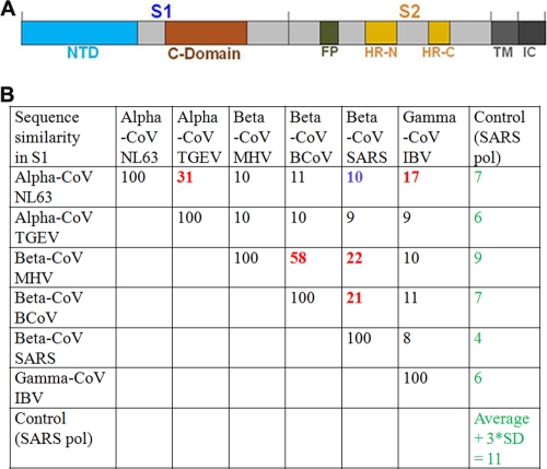
Spike proteins from three coronavirus genera or groups. (A) Schematic representation of coronavirus spike proteins. NTD, N-terminal domain; FP, fusion peptide; HR-N, heptad repeat N; HR-C, heptad repeat C; TM, transmembrane anchor; IC, intracellular tail. (B) Sequence similarities among S1 subunits from representative CoVs (coronaviruses). NL63, human NL63 coronavirus strain Amsterdam I; TGEV, porcine transmissible gastroenteritis virus strain Purdue; MHV, mouse hepatitis coronavirus strain A59; BCoV, bovine coronavirus strain ENT; SARS, SARS coronavirus strain Tor2; IBV, avian infectious bronchitis virus strain M41; SARS pol, SARS polymerase strain Tor02; SD, standard deviation. Alpha-, beta-, and gamma-CoVs can also be referred to as group 1, group 2, and group 3 coronaviruses, respectively. Sequence similarities were calculated using ClusterW (1).
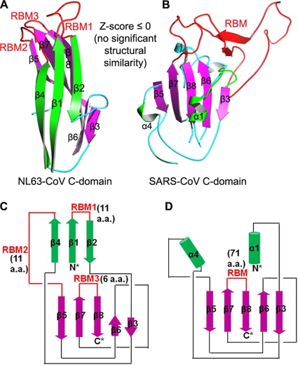
Structures of alpha-coronavirus NL63-CoV and beta-coronavirus SARS-CoV C-domains. (A) Crystal structure of NL63-CoV C-domain (PDB identification no. 3KBH). (B) Crystal structure of SARS-CoV C-domain (PDB identification no. 2AJF). Structural similarity Z-scores were calculated using the DALI server (2). (C) Topology of NL63-CoV C-domain. (D) Topology of SARS-CoV C-domain. RBM, receptor-binding motif.
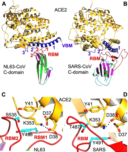
Receptor binding by alpha-coronavirus NL63-CoV and beta-coronavirus SARS-CoV C-domains. (A) Crystal structure of NL63-CoV C-domain complexed with human ACE2 (PDB identification no. 3KBH). (B) Crystal structure of SARS-CoV C-domain complexed with human ACE2 (PDB identification no. 2AJF). (C) A hydrophobic tunnel structure at the NL63-CoV/ACE2 interface, comprising residues Tyr41 and Asp37 from ACE2 and Ser535 and Tyr498 from the NL63-CoV C-domain. (D) A hydrophobic tunnel structure at the SARS-CoV/ACE2 interface, comprising residues Tyr41 and Asp37 from ACE2 and Thr487 and Tyr491 from the SARS-CoV C-domain. The hydrophobic tunnels in panels C and D bury a salt bridge between Lys353 and Asp38 from ACE2. ACE2, angiotensin-converting enzyme 2; VBM, virus-binding motif.
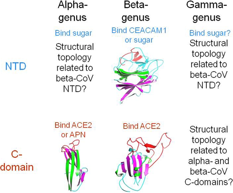
Summary of structure, function, and evolution of coronavirus S1 domains. APN, aminopeptidase N; CEACAM1, carcinoembryonic antigen-related cell adhesion molecule.
References
-
- Li F, Li WH, Farzan M, Harrison SC. 2005. Structure of SARS coronavirus spike receptor-binding domain complexed with receptor. Science 309:1864–1868 - PubMed
Publication types
MeSH terms
Substances
Grants and funding
LinkOut - more resources
Full Text Sources
Other Literature Sources
