. 2012 Mar;86(6):3219-29.
doi: 10.1128/JVI.06712-11. Epub 2011 Dec 28.

Contribution of intra- and interhost dynamics to norovirus evolution

Affiliations

Contribution of intra- and interhost dynamics to norovirus evolution

Rowena A Bull et al. J Virol. 2012 Mar.

Abstract

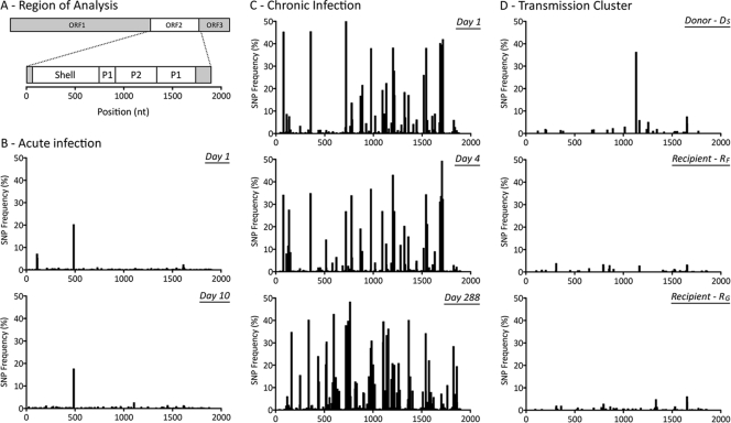
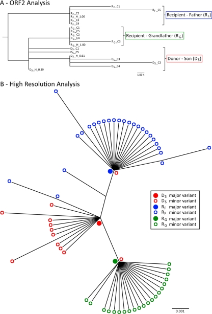
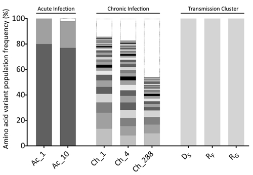
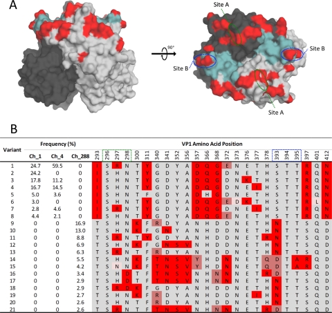
Norovirus (NoV) is an emerging RNA virus that has been associated with global epidemics of gastroenteritis. Each global epidemic arises with the emergence of novel antigenic variants. While the majority of NoV infections are mild and self-limiting, in the young, elderly, and immunocompromised, severe and prolonged illness can result. As yet, there is no vaccine or therapeutic treatment to prevent or control infection. In order to design effective control strategies, it is important to understand the mechanisms and source of the new antigenic variants. In this study, we used next-generation sequencing (NGS) technology to investigate genetic diversification in three contexts: the impact of a NoV transmission event on viral diversity and the contribution to diversity of intrahost evolution over both a short period of time (10 days), in accordance with a typical acute NoV infection, and a prolonged period of time (288 days), as observed for NoV chronic infections of immunocompromised individuals. Investigations of the transmission event revealed that minor variants at frequencies as low as 0.01% were successfully transmitted, indicating that transmission is an important source of diversity at the interhost level of NoV evolution. Our results also suggest that chronically infected immunocompromised subjects represent a potential reservoir for the emergence of new viral variants. In contrast, in a typical acute NoV infection, the viral population was highly homogenous and relatively stable. These results indicate that the evolution of NoV occurs through multiple mechanisms.


Figures

Fig 1
Fig 1
Phylogenetic comparison of the GII.4 ORF2 nucleotide sequence isolated longitudinally from subjects with acute and chronic NoV infections. The full-length ORF2 consensus sequences, determined by bulk sequencing, were generated at two time points (ranging 10 days) for the subject with acute infection (green) and at three time points (ranging 288 days) for the subject with chronic infection (blue). In addition, ORF2 sequences from GII.4 epidemic variants detected in NSW, Australia, during 2006 to 2011 and reference sequences derived from GenBank were included in the analysis (n = 248). Each major GII.4 clade was described previously (59), except for the 2009 and 2010 GII.4 variants, which recently emerged. Both subjects were infected with variants that clustered within the GII.4 2006b variant clade (red). For the subject with acute infection, the consensus sequence was identical at both time points. For the subject with chronic infection, the ORF2 sequence at the third time point differed by 4.1% compared to the sequence from the first two time points. The tree shows that subject Ch was persistently infected with the same variant and not reinfected with another circulating 2006b variant. The distance scale represents the number of nucleotide substitutions per position.
Fig 2
Fig 2
Distribution of single nucleotide polymorphisms (SNPs) detected from the 3′ end of ORF1 to the 5′ end of ORF3. (A) The NoV genomic region analyzed and domains of interest within ORF2, which are shown across the x axes of panels B to D. (B) Distribution of SNPs for subject Ac (acute infection), measured 9 days apart. (C) Distribution of SNPs for subject Ch (chronic infection) at days 1, 4, and 288. (D) Distribution of SNPs for the transmission cluster, involving three acutely infected family members. The donor (subject DS) transmitted the virus to two recipients, subjects RF and RG. A single sample was collected from each infected subject at each time point during the acute stage. The distribution of SNPs, portrayed as a percentage of the viral population, indicates that intrahost viral populations were homogenous for the four acutely infected subjects, with only a few prevalent SNPs (>10%) (B and D). In contrast, subject Ch (C) presented a heterogeneous intrahost population over the course of the infection. Multiple SNPs with a frequency of >10% in the viral population were distributed across the entire length analyzed.
Fig 3
Fig 3
Phylogenetic analysis of sequences from the NoV transmission cluster. (A) Phylogenetic tree of the full-length ORF2 sequences generated from reassembled short NGS reads and from cloning. Sequences are labeled first by their subject name, followed by whether they were generated by cloning (C) or NGS haplotype reconstructions (H). The haplotype frequency is also included at the end of the name. In this cluster, the donor (subject DS) had two closely related variants that were present at high frequencies (38 and 61%) and were identified by both NGS and cloning. However, neither of these two donor variants were found in the viral populations of the two recipients, and each subject's sequences clustered separately. The distance scale represents the number of nucleotide substitutions per position. (B) High-resolution phylogenetic analysis of a region spanning the 3′-terminal 171 nucleotides of ORF2 to the 5′-terminal 133 nucleotides of ORF3. This analysis was performed with NGS data and revealed substantial interhost diversity. For each subject (subjects DS, RF, and RG), the major variant was located at the node of the branch, with minor variants branching from it, indicating that the minor variants had evolved from the dominant variant. In addition, each recipient's major variant was found to be identical to a unique minor variant (<0.01%) isolated from the donor (subject DS). The donor's major variant was not identified in any of the recipient variants at frequencies as low as 0.01%. Filled circles represent major variants within a population, and open circles represent minor variants. Red indicates the donor son (subject DS), while blue represents the recipient father (subject RF) and green represents the recipient grandfather (subject RG). The distance scale represents the number of nucleotide substitutions per position.
Fig 4
Fig 4
Comparison of the intrahost distributions of NoV variants in all subjects. Full-length NoV variants of the ORF2 region were reassembled from NGS reads and translated into amino acid sequences, and each unique variant is represented by alternate gray shading. The histogram shows the frequency distribution of unique NoV variants in each sample analyzed. Low-frequency variants, with an estimated frequency of occurrence below the detection threshold (2%), are indicated by black dotted lines. For the subject with acute infection (subject Ac), only two variants were detected, with frequencies of occurrence of ∼79% and ∼20%, respectively. These variants remained stable over the 9 days of infection. For the subject with chronic infection (subject Ch), no dominant variant was observed. Instead, a distribution of low-frequency variants coexisted, and their prevalences varied over the course of the infection. A single variant was identified in each subject within the transmission cluster cohort (subjects DS, RF, and RG).
Fig 5
Fig 5
Analysis of amino acid variants in the P2 region for subject Ch (chronic NoV infection). (A) Positions of evolving sites (highlighted in red) on the surface of a P2 domain dimer. Each P2 monomer is distinguished by light or dark gray shading. Key antibody binding sites (site A and site B) as well as the histo-blood group antigen (HBGA) binding pocket (highlighted in aqua) are shown. Two orientations are provided: a side view (left) and a top view (right). (B) Amino acid sequence of each NoV variant at 24 amino acid sites, 22 of which were identified as being evolving and polymorphic. The frequency of each variant at days 1, 4, and 288 is provided for subject Ch. Residues that varied between subjects and over time are highlighted by shades of red.

References

    1. Allen DJ, Gray JJ, Gallimore CI, Xerry J, Iturriza-Gomara M. 2008. Analysis of amino acid variation in the P2 domain of the GII-4 norovirus VP1 protein reveals putative variant-specific epitopes. PLoS One 3:e1485. - PMC - PubMed
    1. Allen DJ, et al. 2009. Characterisation of a GII-4 norovirus variant-specific surface-exposed site involved in antibody binding. Virol. J. 6:150. - PMC - PubMed
    1. Arnold K, Bordoli L, Kopp J, Schwede T. 2006. The SWISS-MODEL workspace: a Web-based environment for protein structure homology modelling. Bioinformatics 22:195–201 - PubMed
    1. Belliot G, et al. 2003. In vitro proteolytic processing of the MD145 norovirus ORF1 nonstructural polyprotein yields stable precursors and products similar to those detected in calicivirus-infected cells. J. Virol. 77:10957–10974 - PMC - PubMed
    1. Bertolotti-Ciarlet A, Crawford SE, Hutson AM, Estes MK. 2003. The 3′ end of Norwalk virus mRNA contains determinants that regulate the expression and stability of the viral capsid protein VP1: a novel function for the VP2 protein. J. Virol. 77:11603–11615 - PMC - PubMed

Publication types