. 2012 Apr;81(4):498-509.
doi: 10.1124/mol.111.076356. Epub 2011 Dec 28.

An inducible cytochrome P450 3A4-dependent vitamin D catabolic pathway

Affiliations

An inducible cytochrome P450 3A4-dependent vitamin D catabolic pathway

Zhican Wang et al. Mol Pharmacol. 2012 Apr.

Abstract

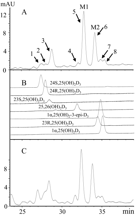
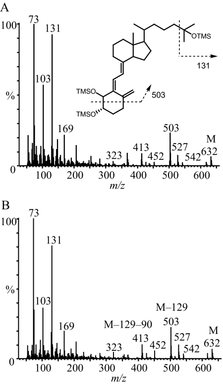
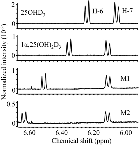
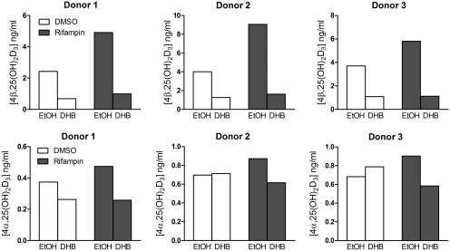
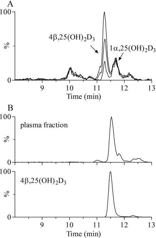
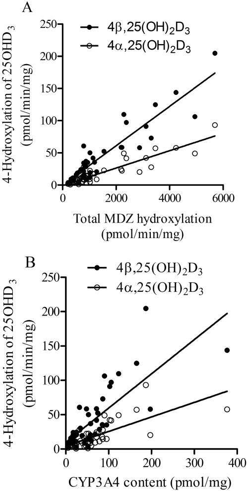
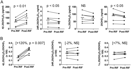
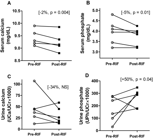
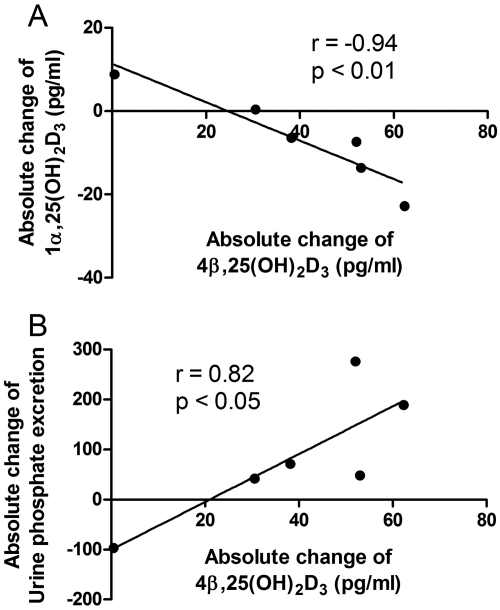
Vitamin D(3) is critical for the regulation of calcium and phosphate homeostasis. In some individuals, mineral homeostasis can be disrupted by long-term therapy with certain antiepileptic drugs and the antimicrobial agent rifampin, resulting in drug-induced osteomalacia, which is attributed to vitamin D deficiency. We now report a novel CYP3A4-dependent pathway, the 4-hydroxylation of 25-hydroxyvitamin D(3) (25OHD(3)), the induction of which may contribute to drug-induced vitamin D deficiency. The metabolism of 25OHD(3) was fully characterized in vitro. CYP3A4 was the predominant source of 25OHD(3) hydroxylation by human liver microsomes, with the formation of 4β,25-dihydroxyvitamin D(3) [4β,25(OH)(2)D(3)] dominating (V(max)/K(m) = 0.85 ml · min(-1) · nmol enzyme(-1)). 4β,25(OH)(2)D(3) was found in human plasma at concentrations comparable to that of 1α,25-dihydroxyvitamin D(3), and its formation rate in a panel of human liver microsomes was strongly correlated with CYP3A4 content and midazolam hydroxylation activity. Formation of 4β,25(OH)(2)D(3) in primary human hepatocytes was induced by rifampin and inhibited by CYP3A4-specific inhibitors. Short-term treatment of healthy volunteers (n = 6) with rifampin selectively induced CYP3A4-dependent 4β,25(OH)(2)D(3), but not CYP24A1-dependent 24R,25-dihydroxyvitamin D(3) formation, and altered systemic mineral homeostasis. Our results suggest that CYP3A4-dependent 25OHD(3) metabolism may play an important role in the regulation of vitamin D(3) in vivo and in the etiology of drug-induced osteomalacia.


Figures

Fig. 1.
Fig. 1.
HPLC profiles of the vitamin D metabolites produced from 25OHD3 by incubation with recombinant CYP3A4 and HLM. A, extracts from 25OHD3 incubation with recombinant CYP3A4. B, seven authentic vitamin D3 metabolites. C, extracts from 25OHD3 incubation with HLM. M1 and M2 denote two major unknown metabolites. mAU, milliabsorbance units.
Fig. 2.
Fig. 2.
Electron impact mass spectra of the trimethylsilyl ether (TMSO) derivatives of the two unknown metabolites, M1 (A) and M2 (B). The metabolites were isolated and purified by HPLC before GC-MS analysis. OTMS, octadecyltrimethoxysilane.
Fig. 3.
Fig. 3.
Identification of M1 and M2 by 1H NMR as 4-hydroxylated 25OHD3 metabolites. The chemical shifts of protons at C-6 and C-7 for 25OHD3, 1α,25(OH)2D3, M1, and M2 were assigned according to literature values (Eguchi and Ikekawa, 1990; Kamao et al., 2004).
Fig. 4.
Fig. 4.
Metabolism of 25OHD3 in primary human hepatocytes. Cells were treated with rifampin (10 μM) or vehicle (0.1% DMSO) for 48 h and then preincubated with DHB (20 μM) or vehicle [0.1% ethanol (EtOH)] to block CYP3A4 activity. Experimental details are shown under Materials and Methods. The culture medium was collected and pooled for LC-MS/MS analysis.
Fig. 5.
Fig. 5.
Detection of 4β,25(OH)2D3 in human plasma. A, LC-MS/MS-based ion current chromatograms of native plasma and native plasma spiked with isolated 4β,25(OH)2D3 (50 and 100 pg). B, ion current chromatograms of HPLC-isolated fractions from native plasma (350 ml) and 4β,25(OH)2D3 standard. MRM was performed by monitoring the transition m/z 574 → 314.
Fig. 6.
Fig. 6.
Correlation between rates of 4-hydroxylation of 25OHD3 and CYP3A4 activity (A)/content (B) in HLM from different liver tissues (n = 42). CYP3A4 activity was expressed as the sum of 1′- and 4-hydroxylation of MDZ, and content was determined by Western blot (Lin et al., 2002).
Fig. 7.
Fig. 7.
Effect of short-term rifampin (RIF) treatment (600 mg. once a day, for 7 days) on the plasma concentrations of dihydroxyvitamin D metabolites (A) and the metabolite to 25OHD3 concentration ratios (B). Individual mean concentrations and ratios at baseline (average of day 0 and day 1, pre-RIF) and after rifampin treatment (average of day 7 and day 8, post-RIF) from six subjects are shown. Bars indicate the mean value for the group. The p value was calculated by paired t test. NS, not significant.
Fig. 8.
Fig. 8.
Effect of short-term rifampin (RIF) treatment on individual serum and urine levels of calcium and phosphate. Serum calcium (A), serum phosphate (B), creatinine-normalized calcium (UCa/UCr × 1000) in spot urine (C), and creatinine-normalized phosphate (UPi/UCr × 1000) in spot urine (D) were measured in six subjects at baseline and after rifampin treatment. For each subject, the averages of two prerifampin measurements (day 0 and day 1) and two post-rifampin measurements (day 7 and day 8) are plotted. NS, not significant.
Fig. 9.
Fig. 9.
Correlation between absolute changes in plasma 4β,25(OH)2D3 concentration with absolute changes in the plasma 1α,25(OH)2D3 level (A) and urinary phosphate excretion (B) from six subjects. The absolute concentration change was calculated as the postrifampin minus prerifampin concentrations. Sample correlation coefficients and corresponding p values were calculated; the null hypothesis that the population correlation is zero was tested with a t test, under the assumption that the population was bivariate normally distributed.
Fig. 10.
Fig. 10.
Proposed metabolic profile of 25OHD3 by recombinant CYP3A4 in vitro. The relative percentage of metabolite/25OHD3 was estimated using HPLC-UV, assuming that each metabolite has the same UV absorption extinction coefficient at 265 nm. In vitro incubation conditions are described under Materials and Methods.

References

    1. Alexander BH, Dimler RJ, Mehltretter CL. (1951) d-Galactosan(1,4)α(1,6): its structure and resistance to periodate oxidation. J Am Chem Soc 73:4658–4659
    1. Araya Z, Hosseinpour F, Bodin K, Wikvall K. (2003) Metabolism of 25-hydroxyvitamin D3 by microsomal and mitochondrial vitamin D3 25-hydroxylases (CYP2D25 and CYP27A1): a novel reaction by CYP27A1. Biochim Biophys Acta 1632:40–47 - PubMed
    1. Bouillon R, Carmeliet G, Verlinden L, van Etten E, Verstuyf A, Luderer HF, Lieben L, Mathieu C, Demay M. (2008) Vitamin D and human health: lessons from vitamin D receptor null mice. Endocr Rev 29:726–776 - PMC - PubMed
    1. Brodie MJ, Boobis AR, Dollery CT, Hillyard CJ, Brown DJ, MacIntyre I, Park BK. (1980) Rifampicin and vitamin D metabolism. Clin Pharmacol Ther 27:810–814 - PubMed
    1. Brodie MJ, Boobis AR, Hillyard CJ, Abeyasekera G, Stevenson JC, MacIntyre I, Park BK. (1982) Effect of rifampicin and isoniazid on vitamin D metabolism. Clin Pharmacol Ther 32:525–530 - PubMed

Publication types

MeSH terms