

Evaluation of cellular phenotypes implicated in immunopathogenesis and monitoring immune reconstitution inflammatory syndrome in HIV/leprosy cases
- PMID: 22205964
- PMCID: PMC3244401
- DOI: 10.1371/journal.pone.0028735
Evaluation of cellular phenotypes implicated in immunopathogenesis and monitoring immune reconstitution inflammatory syndrome in HIV/leprosy cases
Abstract
Background: It is now evident that HAART-associated immunological improvement often leads to a variety of new clinical manifestations, collectively termed immune reconstitution inflammatory syndrome, or IRIS. This phenomenon has already been described in cases of HIV coinfection with Mycobacterium leprae, most of them belonging to the tuberculoid spectrum of leprosy disease, as observed in leprosy reversal reaction (RR). However, the events related to the pathogenesis of this association need to be clarified. This study investigated the immunological profile of HIV/leprosy patients, with special attention to the cellular activation status, to better understand the mechanisms related to IRIS/RR immunopathogenesis, identifying any potential biomarkers for IRIS/RR intercurrence.
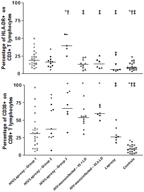
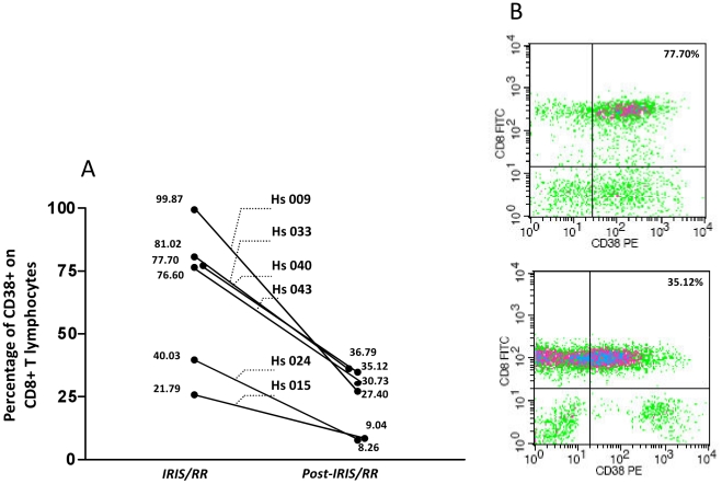
Methods/principal findings: Eighty-five individuals were assessed in this study: HIV/leprosy and HIV-monoinfected patients, grouped according to HIV-viral load levels, leprosy patients without HIV coinfection, and healthy controls. Phenotypes were evaluated by flow cytometry for T cell subsets and immune differentiation/activation markers. As expected, absolute counts of the CD4+ and CD8+ T cells from the HIV-infected individuals changed in relation to those of the leprosy patients and controls. However, there were no significant differences among the groups, whether in the expression of cellular differentiation phenotypes or cellular activation, as reflected by the expression of CD38 and HLA-DR. Six HIV/leprosy patients identified as IRIS/RR were analyzed during IRIS/RR episodes and after prednisone treatment. These patients presented high cellular activation levels regarding the expression of CD38 in CD8+ cells T during IRIS/RR (median: 77,15%), dropping significantly (p<0,05) during post-IRIS/RR moments (median: 29,7%). Furthermore, an increase of cellular activation seems to occur prior to IRIS/RR.
Conclusion/significance: These data suggest CD38 expression in CD8+ T cells interesting tool identifying HIV/leprosy individuals at risk for IRIS/RR. So, a comparative investigation to leprosy patients at RR should be conducted.
Conflict of interest statement
Figures
Similar articles
-
Role of CD8(+) T cells in triggering reversal reaction in HIV/leprosy patients.Immunology. 2013 Sep;140(1):47-60. doi: 10.1111/imm.12108. Immunology. 2013. PMID: 23566249 Free PMC article.
-
Robust Reconstitution of Tuberculosis-Specific Polyfunctional CD4+ T-Cell Responses and Rising Systemic Interleukin 6 in Paradoxical Tuberculosis-Associated Immune Reconstitution Inflammatory Syndrome.Clin Infect Dis. 2016 Mar 15;62(6):795-803. doi: 10.1093/cid/civ978. Epub 2015 Nov 26. Clin Infect Dis. 2016. PMID: 26611774 Free PMC article.
-
Dynamics of T-Lymphocyte Activation Related to Paradoxical Tuberculosis-Associated Immune Reconstitution Inflammatory Syndrome in Persons With Advanced HIV.Front Immunol. 2021 Oct 7;12:757843. doi: 10.3389/fimmu.2021.757843. eCollection 2021. Front Immunol. 2021. PMID: 34691079 Free PMC article.
-
Immune reconstitution inflammatory syndrome in leprosy.Indian J Lepr. 2011 Apr-Jun;83(2):61-70. Indian J Lepr. 2011. PMID: 21972657 Review.
-
Leprosy occurring as immune reconstitution syndrome.Trans R Soc Trop Med Hyg. 2008 Oct;102(10):966-8. doi: 10.1016/j.trstmh.2008.06.003. Epub 2008 Jul 18. Trans R Soc Trop Med Hyg. 2008. PMID: 18639911 Review.
Cited by
-
Leprosy As a Complex Infection: Breakdown of the Th1 and Th2 Immune Paradigm in the Immunopathogenesis of the Disease.Front Immunol. 2017 Nov 28;8:1635. doi: 10.3389/fimmu.2017.01635. eCollection 2017. Front Immunol. 2017. PMID: 29234318 Free PMC article. Review.
-
Role of CD8(+) T cells in triggering reversal reaction in HIV/leprosy patients.Immunology. 2013 Sep;140(1):47-60. doi: 10.1111/imm.12108. Immunology. 2013. PMID: 23566249 Free PMC article.
-
T Cell Activation and Cytokine Profile of Tuberculosis and HIV-Positive Individuals during Antituberculous Treatment and Efavirenz-Based Regimens.PLoS One. 2013 Jun 19;8(6):e66095. doi: 10.1371/journal.pone.0066095. Print 2013. PLoS One. 2013. PMID: 23840403 Free PMC article. Clinical Trial.
-
Understanding the type 1 reactional state for early diagnosis and treatment: a way to avoid disability in leprosy.An Bras Dermatol. 2013 Sep-Oct;88(5):787-92. doi: 10.1590/abd1806-4841.20132004. An Bras Dermatol. 2013. PMID: 24173185 Free PMC article. Review.
-
Immune recovery uveitis in Whipple's disease: an unusual ocular presentation.J Ophthalmic Inflamm Infect. 2024 Feb 13;14(1):10. doi: 10.1186/s12348-024-00390-5. J Ophthalmic Inflamm Infect. 2024. PMID: 38347376 Free PMC article.
References
-
- Hogg RS, O'Shaughnessy MV, Gataric N, Yip B, Craib K, et al. Decline in deaths from AIDS due to new antirretrovirals. Lancet. 1997;349:1294. - PubMed
-
- Palella FJ, Jr, Delaney KM, Moorman AC, Loveless MO, Fuhrer J, et al. Declining morbidity and mortality among patients with advanced human immunodeficiency virus infection. HIV Outpatient Study Investigators. N Engl J Med. 1998;338:853–860. - PubMed
-
- Jehle AW, Khanna N, Sigle JP, Glatz-Krieger K, Battegay M, et al. Acute renal failure on immune reconstitution in an HIV-positive patient with miliary tuberculosis. Clin Infect Dis. 2004;38:e32–35. - PubMed
-
- Race EM, Adelson-Mitty J, Kriegel GR, Barlam TF, Reimann KA, et al. Focal mycobacterial lymphadenitis following initiation of protease-inhibitor therapy in patients with advanced HIV-1 disease. Lancet. 1998;351:252–255. - PubMed
-
- Koval CE, Gigliotti F, Nevins D, Demeter LM. Immune reconstitution syndrome after successful treatment of Pneumocystis carinii pneumonia in a man with human immunodeficiency virus type 1 infection. Clin Infect Dis. 2002;35:491–493. - PubMed
Publication types
MeSH terms
Substances
LinkOut - more resources
Full Text Sources
Medical
Research Materials
