. 2011;6(12):e28780.
doi: 10.1371/journal.pone.0028780. Epub 2011 Dec 21.

Selective cytotoxicity against human osteosarcoma cells by a novel synthetic C-1 analogue of 7-deoxypancratistatin is potentiated by curcumin

Affiliations

Selective cytotoxicity against human osteosarcoma cells by a novel synthetic C-1 analogue of 7-deoxypancratistatin is potentiated by curcumin

Dennis Ma et al. PLoS One. 2011.

Abstract

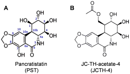
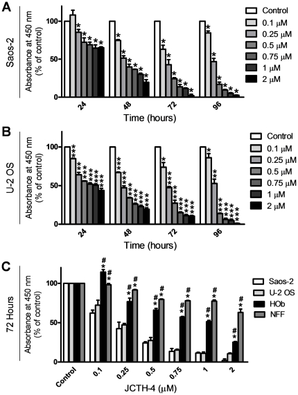
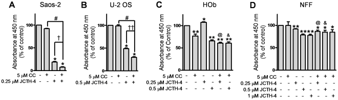
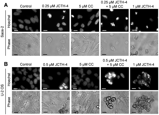
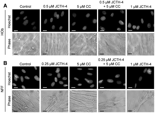
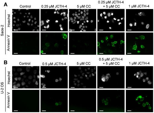
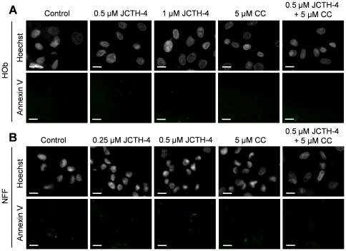
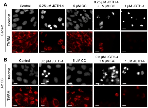
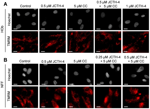
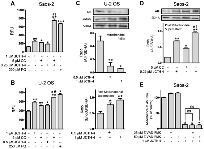
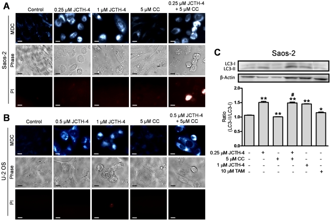
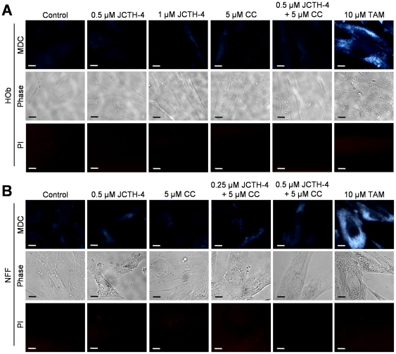
The natural compound pancratistatin (PST) is a non-genotoxic inducer of apoptosis in a variety of cancers. It exhibits cancer selectivity as non-cancerous cells are markedly less sensitive to PST. Nonetheless, PST is not readily synthesized and is present in very low quantities in its natural source to be applied clinically. We have previously synthesized and evaluated several synthetic analogues of 7-deoxypancratistatin, and found that JC-TH-acetate-4 (JCTH-4), a C-1 acetoxymethyl analogue, possessed similar apoptosis inducing activity compared to PST. In this study, notoriously chemoresistant osteosarcoma (OS) cells (Saos-2, U-2 OS) were substantially susceptible to JCTH-4-induced apoptosis through mitochondrial targeting; JCTH-4 induced collapse of mitochondrial membrane potential (MMP), increased reactive oxygen species (ROS) production in isolated mitochondria, and caused release of apoptosis inducing factor (AIF) and endonuclease G (EndoG) from isolated mitochondria. Furthermore, JCTH-4 selectively induced autophagy in OS cells. Additionally, we investigated the combinatory effect of JCTH-4 with the natural compound curcumin (CC), a compound found in turmeric spice, previously shown to possess antiproliferative properties. CC alone had no observable effect on Saos-2 and U-2 OS cells. However, when present with JCTH-4, CC was able to enhance the cytotoxicity of JCTH-4 selectively in OS cells. Such cytotoxicity by JCTH-4 alone and in combination with CC was not observed in normal human osteoblasts (HOb) and normal human fetal fibroblasts (NFF). Therefore, this report illustrates a new window in combination therapy, utilizing a novel synthetic analogue of PST with the natural compound CC, for the treatment of OS.


Conflict of interest statement

Competing Interests: The authors have read the journal's policy and have the following conflict: Dr. Pandey serves on the editorial board for PLoS ONE. This does not alter the authors' adherence to all the PLoS ONE policies on sharing data and materials.

Figures

Figure 1
Figure 1. Comparison of chemical structures.
Structures of (A) Pancratistatin (PST) and (B) JC-TH-acetate-4 (JCTH-4).
Figure 2
Figure 2. JCTH-4 causes selective cytotoxicity in OS cells in a time and dose-dependent manner.
Effect of JCTH-4 on cellular viability of OS cells was determined by the WST-1 based colorimetric assay. (A) Saos-2 and (B) U-2 OS cells were treated with JCTH-4 and the WST-1 reagent was used to quantify cell viability. Absorbance was read at 450 nm and expressed as a percent of the control (Me2SO). Values are expressed as mean ± SD from quadruplicates of 3 independent experiments. *p<0.05, **p<0.01, ***p<0.001 versus solvent control (Me2SO). (C) Effect on cellular viability of HOb and NFF cells treated with JCTH-4 compared to Saos-2 and U-2 OS cells after 72 hours. The WST-1 reagent was used to quantify cellular viability. Absorbance was read at 450 nm and expressed as a percent of the solvent control (Me2SO). Values are expressed as mean ± SD from quadruplicates of 3 independent experiments. *p<0.005 versus Saos-2 cells; #p<0.005 versus U-2 OS cells.
Figure 3
Figure 3. CC potentiates the cytotoxicity of JCTH-4 selectively in OS cells.
Effect of JCTH-4 & CC in combination on cellular viability of OS cells was determined by the WST-1 based colorimetric assay. (A) Saos-2 (96 hours), (B) U-2 OS (72 hours), (C) HOb (72 hours), and (D) NFF (72 hours) cells were treated with JCTH-4 and CC and the WST-1 reagent was used to quantify cellular viability. Absorbance was read at 450 nm and expressed as a percent of the solvent control (Me2SO). Values are expressed as mean ± SD from quadruplicates of 3 independent experiments. *p<0.05, **p<0.01, versus solvent control (Me2SO); †p<0.001 versus 0.25 µM JCTH-4; ††p<0.01 versus 0.5 µM JCTH-4; #p<0.001 versus 5 µM CC; @p<0.001 versus 0.25 µM JCTH-4+5 µM CC treatment with Saos-2 cells (Figure 3A); &p<0.01 versus 0.5 µM JCTH-4+5 µM CC treatment with U-2 OS cells (Figure 3B).
Figure 4
Figure 4. JCTH-4 alone and in combination with CC yields apoptotic morphology in OS cells.
Nuclear and cellular morphology of (A) Saos-2 cells after 96 hours of treatment and (B) U-2 OS cells after 72 hours of treatment. Cells were treated with JCTH-4, CC, and solvent control (Me2SO). Post treatment, the cells were stained with Hoechst 33342 dye. Corresponding phase micrographs are shown below the Hoechst micrographs. Apoptotic morphology is evident in cells with bright and condensed nuclei accompanied by apoptotic bodies, as well as cell shrinkage and blebbing. Images were taken at 400× magnification on a fluorescent microscope. Scale bar = 15 µm. All images are representative of 3 independent experiments.
Figure 5
Figure 5. JCTH-4 and CC do not yield apoptotic morphology in HOb and NFF cells.
Nuclear and cellular morphology of (A) HOb and (B) NFF cells after 72 hours of treatment. Cells were treated with JCTH-4, CC, and solvent control (Me2SO). Post treatment, the cells were stained with Hoechst 33342 dye. Corresponding phase micrographs are shown below the Hoechst micrographs. Apoptotic morphology is evident in cells with bright and condensed nuclei accompanied by apoptotic bodies, as well as cell shrinkage and blebbing. Images were taken at 400× magnification on a fluorescent microscope. Scale bar = 15 µm. All images are representative of 3 independent experiments.
Figure 6
Figure 6. JCTH-4 alone and in combination with CC induces phosphatidylserine externalization in OS cells.
Annexin V binding to externalized phosphatidylserine was monitored to confirm induction of apoptosis in (A) Saos-2 cells after 96 hours of treatment and (B) U-2 OS cells after 72 hours of treatment with the indicated concentrations of JCTH-4, CC, and solvent control (Me2SO). Cells were also stained with Hoechst dye. Images were taken at 400× magnification on a fluorescent microscope. Green fluorescence is indicative of Annexin V binding to externalized phosphatidylserine of the plasma membrane. Scale bar = 15 µm. All images are representative of 3 independent experiments.
Figure 7
Figure 7. JCTH-4 alone and with CC does not induce phosphatidylserine externalization in HOb and NFF cells.
Annexin V binding to externalized phosphatidylserine was monitored to confirm that apoptosis was not induced in (A) HOb and (B) NFF cells after 72 hours of treatment. Cells were treated with the indicated concentrations of JCTH-4, CC and solvent control (Me2SO). Cells were also stained with Hoechst dye. Images were taken at 400× magnification on a fluorescent microscope. Green fluorescence is indicative of Annexin V binding to externalized phosphatidylserine of the plasma membrane. Scale bar = 15 µm. All images are representative of 3 independent experiments.
Figure 8
Figure 8. JCTH-4 dissipates MMP alone and in combination with CC in OS cells.
Effect of JCTH-4 and CC on MMP in (A) Saos-2 cells after 96 hours of treatment and (B) U-2 OS cells after 72 hours of treatment was examined by TMRM staining. Cells were grown on coverslips, treated with the indicated concentrations of JCTH-4, CC, and solvent control (Me2SO) and stained with TMRM and Hoechst dye. Images were taken at 400× magnification on a fluorescent microscope. Red fluorescent punctuate marks are indicative of mitochondria with intact MMP. Scale bar = 15 µm. All images are representative of 3 independent experiments.
Figure 9
Figure 9. JCTH-4 and CC do not dissipate MMP in HOb and NFF cells.
Effect of JCTH-4 and CC on MMP in (A) HOb and (B) NFF cells after 72 hours of treatment was examined by TMRM staining. Cells were grown on coverslips, treated with the indicated concentrations of JCTH-4, CC, and solvent control (Me2SO), and stained with TMRM and Hoechst dye. Images were taken at 400× magnification on a fluorescent microscope. Red fluorescent punctuate marks are indicative of mitochondria with intact MMP. Scale bar = 15 µm. All images are representative of 3 independent experiments.
Figure 10
Figure 10. JCTH-4 directly causes mitochondrial ROS production and release of apoptogenic factors independent of caspases.
(A) Saos-2 and (B) U-2 OS isolated mitochondria were treated directly with JCTH-4, CC, PQ, and solvent control (Me2SO), and incubated with Amplex Red and horseradish peroxidase for 2 hours. Subsequently, fluorescence readings were taken at Ex. 560 nm and Em. 590 nm and expressed as relative fluorescence units (RFU). Statistics were performed using GraphPad Prism version 5.0. Image is representative of 3 independent experiments demonstrating similar trends. Values are expressed as mean ± SD of quadruplicates of 1 independent experiment. *p<0.05, **p<0.01, ***p<0.001 versus solvent control (Me2SO); †p<0.01 versus 0.25 µM JCTH-4; @p<0.01 versus 0.5 µM JCTH-4; #p<0.01 versus 5 µM CC. Isolated mitochondria samples treated directly with JCTH-4, CC, and solvent control (Me2SO) for 2 hours were also centrifuged, producing mitochondrial pellets and post mitochondrial supernatants which were examined for retention and release of apoptogenic factors respectively via western blot analyses; (C) Retention of AIF and release of EndoG by U-2 OS cell mitochondria and (D) release of AIF by Saos-2 cell mitochondria was monitored. Mitochondrial pellets were probed for SDHA to serve as loading controls. Densitometric analyses were performed using ImageJ software and statistics were calculated using GraphPad Prism version 5.0. Image is representative of 3 independent experiments demonstrating similar trends. Values are expressed as mean ± SD of triplicates of one independent experiment. *p<0.01, **p<0.001 versus solvent control (Me2SO); †p<0.01 versus 0.25 µM JCTH-4; #p<0.01 versus 5 µM CC. (E) Saos-2 cells were treated with broad spectrum caspase inhibitor Z-VAD-FMK with and without JCTH-4 for 72 hours. WST-1 reagent was used to quantify cell viability. Absorbance was read at 450 nm and expressed as a percent of solvent control (Me2SO). Values are expressed as mean ± SD from quadruplicates of 3 independent experiments. *p<0.001 versus solvent control (Me2SO); ns = not significant.
Figure 11
Figure 11. JCTH-4 induces autophagy in OS cells alone and with CC.
The presence of autophagic vacuoles in (A) Saos-2 after 96 hours of treatment and (B) U-2 OS cells after 72 hours of treatment with JCTH-4, CC, and solvent control (Me2SO) was determined by MDC staining. Bright blue punctate marks are indicative of autophagic vacuoles. Corresponding phase and PI micrographs are shown below the MDC images. Scale bar = 15 µm. All images are representative of 3 independent experiments. (C) Cell lysates of Saos-2 cells treated with JCTH-4, CC, TAM as positive control, and solvent control (Me2SO) for 72 hours were examined for the conversion of LC3-I to LC3-II by western blot analyses. β-actin was probed to serve as a loading control. Densitometric analyses were done using ImageJ software and statistics were calculated using GraphPad Prism version 5.0. Image is representative of 3 independent experiments demonstrating similar trends. Values are expressed as mean ± SD of triplicates of one independent experiment. *p<0.05, **p<0.01 versus solvent control (Me2SO); #p<0.01 versus 5 µM CC.
Figure 12
Figure 12. JCTH-4 and CC do not induce autophagy in Hob and NFF cells.
MDC staining was used to detect the presence of autophagic vacuoles in (A) HOb and (B) NFF cells after 72 hours of treatment with JCTH-4, CC, and solvent control (Me2SO) at the indicated concentrations. Bright blue punctate marks are indicative of autophagic vacuoles. Corresponding phase and PI micrographs are shown below the MDC images. Scale bar = 15 µm. All images are representative of 3 independent experiments.

Similar articles

Cited by

References

    1. Aggarwal B, Kumar A, Bharti A. Anticancer potential of curcumin: preclinical and clinical studies. Anticancer Res. 2003;23(1A):363–398. - PubMed
    1. Singh S. From exotic spice to modern drug? Cell. 2007;130:765–768. - PubMed
    1. Kuttan R, Bhanumathy P, Nirmala K, George M. Potential anticancer activity of turmeric (Curcuma longa). Cancer Lett. 1985;29(2):197–202. - PubMed
    1. Walters DK, Muff R, Langsam B, Born W, Fuchs B. Cytotoxic effects of curcumin on osteosarcoma cell lines. Invest New Drugs. 2008;26(4):289–97. Epub 2007 Dec 11. - PubMed
    1. Arndt CA, Crist WM. Common musculoskeletal tumors of childhood and adolescence. N Engl J Med. 1999;341:342–352. - PubMed

Publication types

MeSH terms