. 2011;6(12):e28842.
doi: 10.1371/journal.pone.0028842. Epub 2011 Dec 20.

CCL5 neutralization restricts cancer growth and potentiates the targeting of PDGFRβ in colorectal carcinoma

Affiliations

CCL5 neutralization restricts cancer growth and potentiates the targeting of PDGFRβ in colorectal carcinoma

Béatrice Cambien et al. PLoS One. 2011.

Abstract

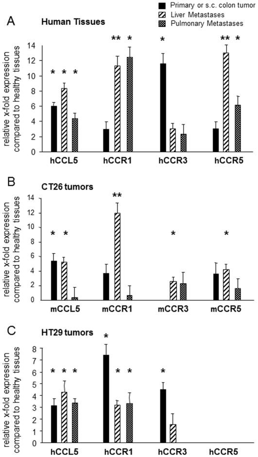
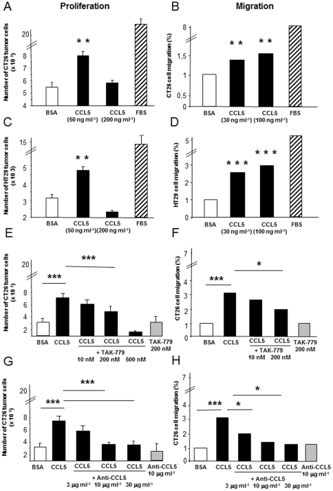
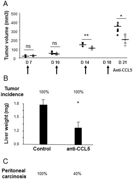
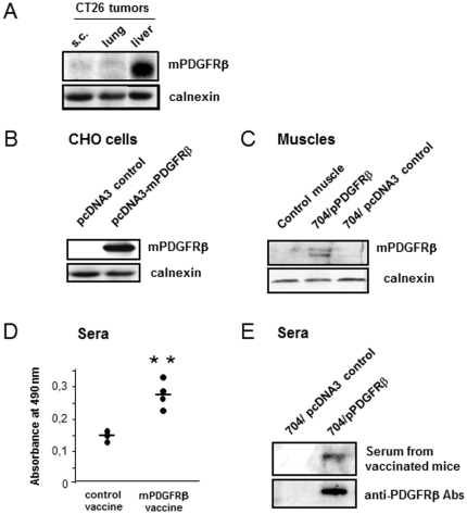
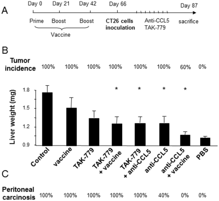
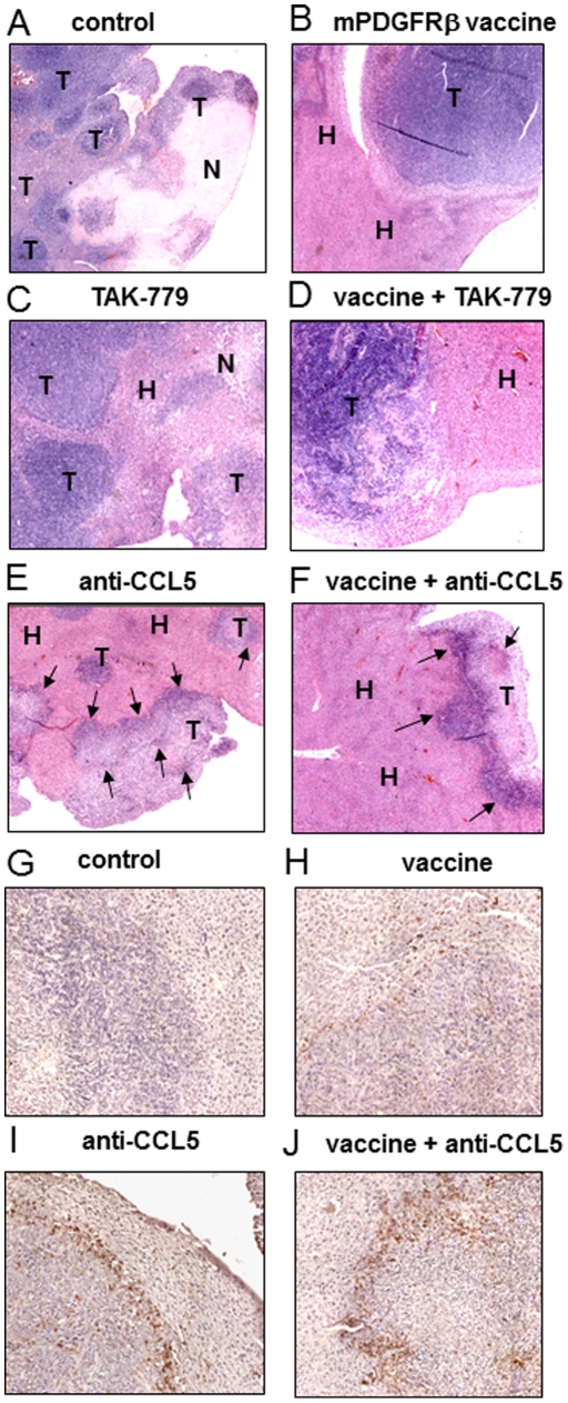
Increased CCL5 levels are markers of an unfavourable outcome in patients with melanoma, breast, cervical, prostate, gastric or pancreatic cancer. Here, we have assessed the role played by CCL5/CCR5 interactions in the development of colon cancer. To do so, we have examined a number of human colorectal carcinoma clinical specimens and found CCL5 and its receptors over-expressed within primary as well as liver and pulmonary metastases of patients compared to healthy tissues. In vitro, CCL5 increased the growth and migratory responses of colon cancer cells from both human and mouse origins. In addition, systemic treatment of mice with CCL5-directed antibodies reduced the extent of development of subcutaneous colon tumors, of liver metastases and of peritoneal carcinosis. Consistently, we found increased numbers of CD45-immunoreactive cells within the stroma of the remaining lesions as well as at the interface with the healthy tissue. In contrast, selective targeting of CCR5 through administration of TAK-779, a CCR5 antagonist, only partially compromised colon cancer progression. Furthermore, CCL5 neutralization rendered the tumors more sensitive to a PDGFRβ-directed strategy in mice, this combination regimen offering the greatest protection against liver metastases and suppressing macroscopic peritoneal carcinosis. Collectively, our data demonstrate the involvement of CCL5 in the pathogenesis of colorectal carcinoma and point to its potential value as a therapeutic target.


Conflict of interest statement

Competing Interests: Bruno Pitard owns stock in In-Cell-Art Co., which develops 704 for vaccine applications. This does not alter the authors' adherence to all the PLoS ONE policies on sharing data and materials.

Figures

Figure 1
Figure 1. Expression of CCL5 and its corresponding CCR1, CCR3, CCR5 receptors in human biopsies and mouse tissues of colorectal carcinoma.
Analysis of levels of expression of human (h) or mouse (m) targets was performed by quantitative RT-PCR in surgical resection pieces of human colorectal carcinoma (n = 10) (A), in experimental subcutaneous tumors (n = 6), liver (n = 6) and lung metastases (n = 6) derived from mouse CT26 cells (B) or derived from human HT29 cells (C) compared with corresponding healthy tissues (normal human, n = 10, and normal mouse colon, n = 6, respectively). The relative levels of expression were calculated using standard curves and expressed as 1/ΔCt. ΔCt values were calculated by substracting Ct of normalizing gene from Ct of target gene, measured in the same RNA preparation. Comparative level of mRNA expression between healthy (X) and metastatic tissues (Y) was calculated using the formula ΔC T Y – ΔC T X and expressed as fold over healthy (2ΔΔC T). Black bars: primary or subcutaneous colon tumors; hatched bars: liver metastases; dotted bars: lung metastases. * p<0.05, ** p<0.01.
Figure 2
Figure 2. CCL5-induced tumor-promoting properties in CRC cells.
(A, C, E, G) Proliferation of the CT26 and HT29 cells was assessed in response to a 5-day treatment with base medium alone (BSA, open bars), with serum-enriched medium (FBS, hatched bars) or with the indicated concentrations of recombinant CCL5 (filled bars), in the presence or in the absence of TAK-779 or anti-CCL5 antibodies (at the indicated concentrations). (B, D, F, H) CRC cells were assayed for chemotaxis in response to base medium alone (BSA, open bars), to serum-enriched medium (FBS, hatched bars) or to the indicated concentrations of recombinant CCL5 (filled bars), in the presence or in the absence of TAK-779 or anti-CCL5 antibodies. Results represent the mean±s.e.m. of six determinations. * p<0.05, ** p<0.01, *** p<0.001.
Figure 3
Figure 3. Protective effect of CCL5 neutralization on the development of colorectal tumors, metastases and peritoneal carcinosis.
(A) Mice subcutaneously-challenged with CT26 cells received four intratumor injections of anti-CCL5 (open dots) or isotype-matched antibodies (filled dots) at the indicated times. Upon sacrifice, the extent of tumor development was assessed by measuring the tumor burdens. (B) Incidence and extent of tumor development within livers of CT26-challenged mice treated with anti-CCL5- or isotype-matched antibodies. (C) Incidence of peritoneal carcinosis expressed as percent of mice bearing carcinosis. (n = 5/group). * p<0.05, ** p<0.01.
Figure 4
Figure 4. Formulation of DNA with 704 polymer increases mPDGFRβ expression level after intramuscular injection and induces a specific immune response.
Western blot analyses of mPDGFRβ expression level (A) in CT26-derived subcutaneous tumors, pulmonary and liver metastases, (B) in pcDNA-3 control (with antisens PCR product) and pcDNA3-mPDGFRβ-transfected CHO cells and (C) in tibialis anterior muscles of Balb/c mice injected once with low-dose (25 µg) of mPDGFRβ encoding plasmid formulated in 704 copolymer (n = 4). (D) Determination of serum levels of mPDGFRβ-specific antibodies by ELISA. Mice were challenged on day 0, and restimulated on day +21, and day +42 with the DNA vaccine (50 µg) or the control vector formulated with 0.3% 704 before being sacrificed two weeks later for serum collection (n = 5). ** p<0.01. (E) Mouse sera were assayed for their ability to react with the band of the purified antigen from the mPDGFRβ-transfected CHO cells by Western blot analysis. Goat anti-mPDGFRβ antibodies from Santa Cruz were used for comparison.
Figure 5
Figure 5. Combining CCL5- and PDGFRβ-directed strategies offers the greatest protection against liver tumors and peritoneal carcinosis.
(A) Mice were challenged on day 0, and restimulated on day +21, and day +42 with the DNA vaccine (50 µg) or the control vector formulated with 0.3% poloxamine 704 before receiving three weeks later an injection of CT26 cells under the liver capsule. Seventy two hours later, mice were treated with CCL5-directed antibodies or with TAK-779 or both, as described in the Materials and Methods section. (B) Incidence and extent of tumor development within livers of CT26-challenged mice, immunized or not against PDGFRβ and treated either with anti-CCL5 antibodies, with TAK-779, or with both blockers. (C) Incidence of peritoneal carcinosis expressed as percent of mice bearing carcinosis. (n = 5/group). * p<0.05, ** p<0.01.
Figure 6
Figure 6. Liver histology at the time of sacrifice.
(A–F) Liver histology was compared on sections of organs from CT26-challenged mice either untreated (A), or immunized with mPDGFRβ vaccine (B), or treated with TAK-779 (C), with TAK-779 plus mPDGFRβ vaccine (D), with anti-CCL5 antibodies (E) or with anti-CCL5 antibodies plus mPDGFRβ vaccine (F). T = liver tumors produced by CT26 cells; N = area of necrosis in a liver tumor; H = hepatocytes; Arrows indicate the presence of dense infiltrates at the interface with the healthy tissue and within lesions. (G–J) CD45-staining reveals immunoreactive leukocytes at the interface tumor-parenchyma and within hepatic lesions of anti-CCL5- (I) and anti-CCL5/PDGFRβ-treated mice (J) but less leukocytes within vaccinated mice (H) and almost no staining in untreated mice (G).

References

    1. Mueller MM, Fusenig NE. Friends or foes - bipolar effects of the tumour stroma in cancer. Nat Rev Cancer. 2004;4(11):839–49. - PubMed
    1. Bhowmick NA, Neilson EG, Moses HL. Stromal fibroblasts in cancer initiation and progression. Nature. 2004;432(7015):332–7. - PMC - PubMed
    1. Tlsty TD, Coussens LM. Tumor stroma and regulation of cancer development. Annu Rev Pathol. 2006;1:119–50. - PubMed
    1. Ito M, Yoshida K, Kyo E, Ayhan A, Nakayama H, et al. Expression of several growth factors and their receptor genes in human colon carcinomas. Virchows Arch B Cell Pathol Incl Mol Pathol. 1990;59(3):173–8. - PubMed
    1. De Jong KP, Stellema R, Karrenbeld A, Koudstaal J, Gouw AS, et al. Clinical relevance of transforming growth factor alpha, epidermal growth factor receptor, p53, and Ki67 in colorectal liver metastases and corresponding primary tumors. Hepatology. 1998;28(4):971–9. - PubMed

Publication types

MeSH terms