

Suppression and regression of choroidal neovascularization in mice by a novel CCR2 antagonist, INCB3344
- PMID: 22205983
- PMCID: PMC3242774
- DOI: 10.1371/journal.pone.0028933
Suppression and regression of choroidal neovascularization in mice by a novel CCR2 antagonist, INCB3344
Abstract
Purpose: To investigate the effect of an intravitreally administered CCR2 antagonist, INCB3344, on a mouse model of choroidal neovascularization (CNV).
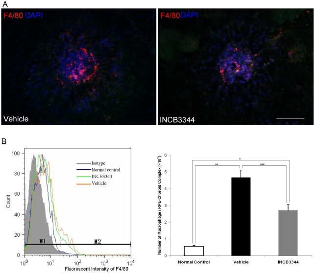
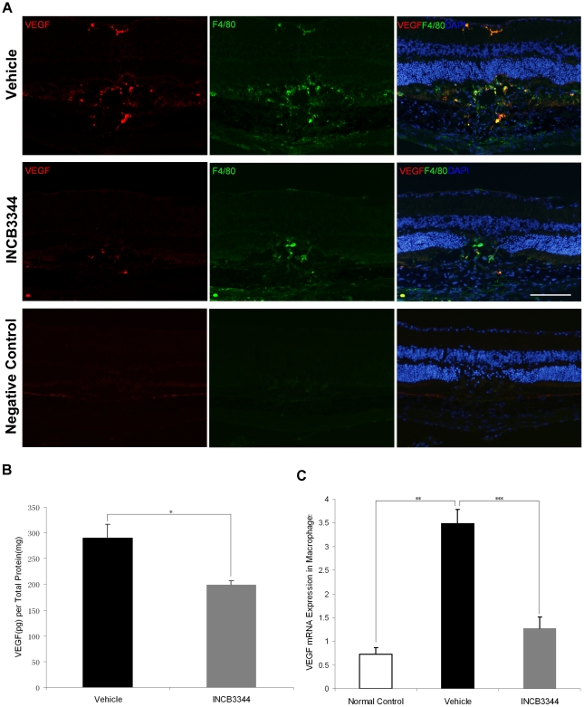
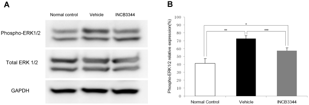
Methods: CNV was induced by laser photocoagulation on Day 0 in wild type mice. INCB3344 or vehicle was administered intravitreally immediately after laser application. On Day 14, CNV areas were measured on retinal pigment epithelium (RPE)-choroid flat mounts and histopathologic examination was performed on 7 µm-thick sections. Macrophage infiltration was evaluated by immunohistochemistry on RPE-choroid flat mounts and quantified by flow cytometry on Day 3. Expression of vascular endothelial growth factor (VEGF) protein in RPE-choroid tissue was examined by immunohistochemistry and ELISA, VEGF mRNA in sorted macrophages in RPE-choroid tissue was examine by real-time PCR and expression of phosphorylated extracellular signal-regulated kinase (p-ERK 1/2) in RPE-choroid tissue was measured by Western blot analysis on Day 3. We also evaluated the efficacy of intravitreal INCB3344 to spontaneous CNV detected in Cu, Zn-superoxide dismutase (SOD1) deficient mice. Changes in CNV size were assessed between pre- and 1week post-INCB3344 or vehicle administration in fundus photography and fluorescence angiography (FA).
Results: The mean CNV area in INCB3344-treated mice decreased by 42.4% compared with the vehicle-treated control mice (p<0.001). INCB3344 treatment significantly inhibited macrophage infiltration into the laser-irradiated area (p<0.001), and suppressed the expression of VEGF protein (p = 0.012), VEGF mRNA in infiltrating macrophages (p<0.001) and the phosphorylation of ERK1/2 (p<0.001). The area of spontaneous CNV in Sod1⁻/⁻ mice regressed by 70.35% in INCB3344-treated animals while no change was detected in vehicle-treated control mice (p<0.001).
Conclusions: INCB3344 both inhibits newly forming CNV and regresses established CNV. Controlling inflammation by suppressing macrophage infiltration and angiogenic ability via the CCR-2/MCP-1 signal may be a useful therapeutic strategy for treating CNV associated with age-related macular degeneration.
Conflict of interest statement
Figures


Similar articles
-
Suppression of experimental choroidal neovascularization by curcumin in mice.PLoS One. 2012;7(12):e53329. doi: 10.1371/journal.pone.0053329. Epub 2012 Dec 28. PLoS One. 2012. PMID: 23285282 Free PMC article.
-
Lactic Acid Upregulates VEGF Expression in Macrophages and Facilitates Choroidal Neovascularization.Invest Ophthalmol Vis Sci. 2018 Jul 2;59(8):3747-3754. doi: 10.1167/iovs.18-23892. Invest Ophthalmol Vis Sci. 2018. PMID: 30046816
-
Antiangiogenic effects of tivozanib, an oral VEGF receptor tyrosine kinase inhibitor, on experimental choroidal neovascularization in mice.Exp Eye Res. 2013 Jul;112:125-33. doi: 10.1016/j.exer.2013.05.006. Epub 2013 May 20. Exp Eye Res. 2013. PMID: 23701975
-
Anti-angiogenic effect of ALS-L1023, an extract of Melissa officinalis L., on experimental choroidal neovascularization in mice.Clin Exp Ophthalmol. 2016 Jan-Feb;44(1):43-51. doi: 10.1111/ceo.12583. Epub 2015 Oct 1. Clin Exp Ophthalmol. 2016. PMID: 26221970
-
Blockade of vascular adhesion protein-1 attenuates choroidal neovascularization.Mol Vis. 2012;18:593-600. Epub 2012 Mar 2. Mol Vis. 2012. PMID: 22419852 Free PMC article.
Cited by
-
Age-dependent changes in FasL (CD95L) modulate macrophage function in a model of age-related macular degeneration.Invest Ophthalmol Vis Sci. 2013 Aug 7;54(8):5321-31. doi: 10.1167/iovs.13-12122. Invest Ophthalmol Vis Sci. 2013. PMID: 23821188 Free PMC article.
-
ERK1/2 activation is a therapeutic target in age-related macular degeneration.Proc Natl Acad Sci U S A. 2012 Aug 21;109(34):13781-6. doi: 10.1073/pnas.1206494109. Epub 2012 Aug 6. Proc Natl Acad Sci U S A. 2012. PMID: 22869729 Free PMC article.
-
Promiscuous Chemokine Antagonist (BKT130) Suppresses Laser-Induced Choroidal Neovascularization by Inhibition of Monocyte Recruitment.J Immunol Res. 2019 Aug 5;2019:8535273. doi: 10.1155/2019/8535273. eCollection 2019. J Immunol Res. 2019. PMID: 31467935 Free PMC article.
-
VEGF-production by CCR2-dependent macrophages contributes to laser-induced choroidal neovascularization.PLoS One. 2014 Apr 8;9(4):e94313. doi: 10.1371/journal.pone.0094313. eCollection 2014. PLoS One. 2014. PMID: 24714223 Free PMC article.
-
CCL2, CCR2 Gene Variants and CCL2, CCR2 Serum Levels Association with Age-Related Macular Degeneration.Life (Basel). 2022 Jul 12;12(7):1038. doi: 10.3390/life12071038. Life (Basel). 2022. PMID: 35888126 Free PMC article.
References
-
- Fine SL, Berger JW, Maguire MG, Ho AC. Age-related macular degeneration. N Engl J Med. 2000;342:483–492. - PubMed
-
- Yamada K, Sakurai E, Itaya M, Yamasaki S, Ogura Y. Inhibition of laser-induced choroidal neovascularization by atorvastatin by downregulation of monocyte chemotactic protein-1 synthesis in mice. Invest Ophthalmol Vis Sci. 2007;48:1839–1843. - PubMed
-
- Grossniklaus HE, Green WR. Choroidal neovascularization. Am J Ophthalmol. 2004;137:496–503. - PubMed
-
- Grossniklaus HE, Ling JX, Wallace TM, Dithmar S, Lawson DH, et al. Macrophage and retinal pigment epithelium expression of angiogenic cytokines in choroidal neovascularization. Mol Vis. 2002;8:119–126. - PubMed
Publication types
MeSH terms
Substances
LinkOut - more resources
Full Text Sources
Other Literature Sources
Miscellaneous
