. 2011;6(12):e29099.
doi: 10.1371/journal.pone.0029099. Epub 2011 Dec 20.

Mechanisms of MEOX1 and MEOX2 regulation of the cyclin dependent kinase inhibitors p21 and p16 in vascular endothelial cells

Affiliations

Mechanisms of MEOX1 and MEOX2 regulation of the cyclin dependent kinase inhibitors p21 and p16 in vascular endothelial cells

Josette M Douville et al. PLoS One. 2011.

Abstract

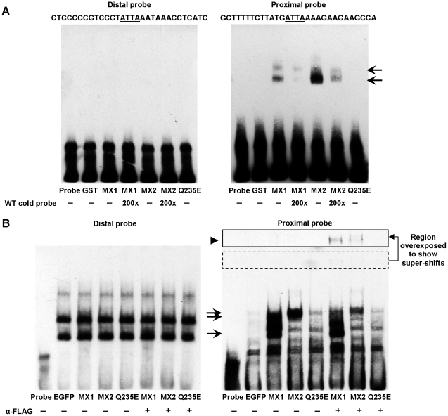
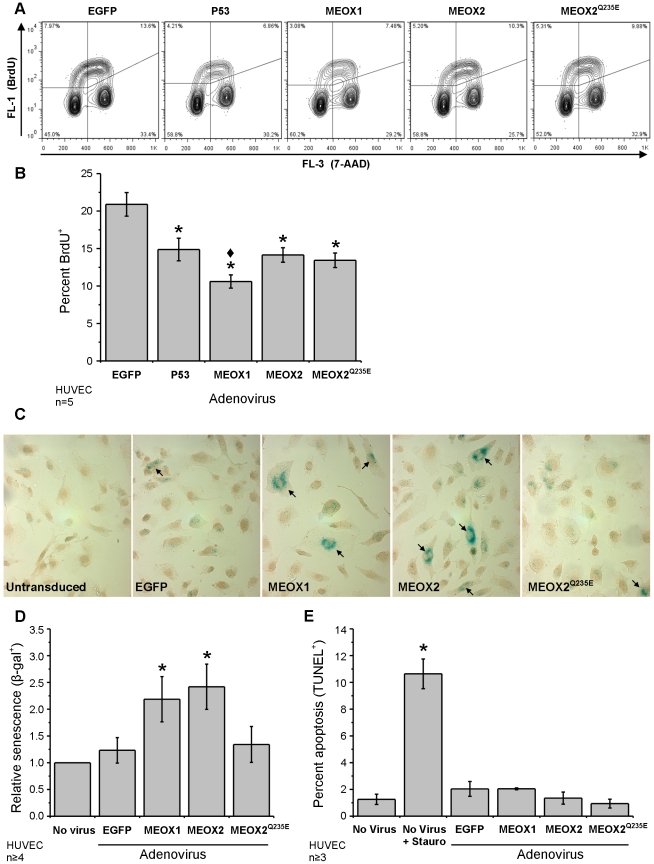
Senescence, the state of permanent cell cycle arrest, has been associated with endothelial cell dysfunction and atherosclerosis. The cyclin dependent kinase inhibitors p21(CIP1/WAF1) and p16(INK4a) govern the G(1)/S cell cycle checkpoint and are essential for determining whether a cell enters into an arrested state. The homeodomain transcription factor MEOX2 is an important regulator of vascular cell proliferation and is a direct transcriptional activator of both p21(CIP1/WAF1) and p16(INK4a). MEOX1 and MEOX2 have been shown to be partially functionally redundant during development, suggesting that they regulate similar target genes in vivo. We compared the ability of MEOX1 and MEOX2 to activate p21(CIP1/WAF1) and p16(INK4a) expression and induce endothelial cell cycle arrest. Our results demonstrate for the first time that MEOX1 regulates the MEOX2 target genes p21(CIP1/WAF1) and p16(INK4a). In addition, increased expression of either of the MEOX homeodomain transcription factors leads to cell cycle arrest and endothelial cell senescence. Furthermore, we show that the mechanism of transcriptional activation of these cyclin dependent kinase inhibitor genes by MEOX1 and MEOX2 is distinct. MEOX1 and MEOX2 activate p16(INK4a) in a DNA binding dependent manner, whereas they induce p21(CIP1/WAF1) in a DNA binding independent manner.


Conflict of interest statement

Competing Interests: The authors have declared that no competing interests exist.

Figures

Figure 1
Figure 1. Expression and subcellular localization of MEOX1 and MEOX2 proteins.
A) Schematic representation of the MEOX1 and MEOX2 protein constructs used in this paper. The alignment lists the percentage of similar and identical amino acids conserved between human MEOX1 and MEOX2 in the different functional protein domains. B) Representative western blot displaying the relative level of MEOX1 (MX1), MEOX2 (MX2), DNA binding deficient MEOX1Q220E (Q220E), DNA binding deficient MEOX2Q235E (Q235E) and homeodomain deleted MEOX2K195_K245del (K195_K245del) protein expression in HEK293 cells 48 hours after transfection. The N-terminally tagged MEOX proteins were detected using an anti-FLAG antibody and α-tubulin was used as a loading control. The empty expression vector was used as a negative control (Ctrl). C) A representative western blot demonstrating the subcellular localization of different MEOX proteins in HEK293 cells, 48 hours after transfection. α-tubulin was used as a cytoplasmic (C) marker and lamin A/C was used as nuclear (N) marker. D) Representative fluorescent immunocytochemistry showing the localization and level of expression of the MEOX proteins in HUVECs 48 hours after adenoviral transduction at a multiplicity of infection of 250. The N-terminally tagged MEOX proteins were detected using an anti-FLAG antibody (green) and nuclei were stained with propidium iodide (red). Enhanced green fluorescent protein (EGFP) was used as a control for adenoviral infection. Arrows indicate cytoplasmic staining and arrowheads indicate punctate nuclear aggregates. Scale bar represents 20 µm.
Figure 2
Figure 2. MEOX1 and MEOX2 activate expression of p21CIP1/WAF1 mRNA and protein in endothelial cells.
A) Relative level of endogenous p21CIP1/WAF1 mRNA compared to EGFP transduced HUVECs. Total RNA was isolated from HUVECs 48 hours after adenoviral transduction at a multiplicity of infection (MOI) of 250 and the amount of p21CIP1/WAF1 mRNA was measured by quantitative real-time PCR. Beta-actin mRNA expression was used for inter-sample normalisation. B) A representative western blot showing increased p21CIP1/WAF1 protein in HUVECs expressing ectopic MEOX2 (MX2) or DNA binding mutant MEOX2Q235E (Q235E) but not MEOX1 (MX1). Total protein was isolated from HUVECs 48 hours after adenoviral transduction at 250 MOI. C) Quantification of the relative amount of p21CIP1/WAF1 protein compared to EGFP transduced HUVECs. The intensity of the p21CIP1/WAF1 band was normalized to the actin loading control. D) Activation of the 2272 bp p21CIP1/WAF1 promoter driven luciferase reporter gene by MEOX1 and MEOX2 in HEK293 and HUVECs. * Indicates a statistically significant change (p<0.05) when compared to the empty vector or EGFP control. ♦ Indicates a statistically significant difference (p<0.05) between MEOX1 and MEOX2.
Figure 3
Figure 3. MEOX1 and MEOX2 activate transcription of the p16INK4a gene in HUVECs.
A) Relative level of p16INK4a mRNA in HUVECs. Total RNA was isolated from HUVECs 72 hours after adenoviral transduction at a multiplicity of infection (MOI) of 250 and the amount of p16INK4a mRNA was measured by quantitative real-time PCR and compared to EGFP transduced HUVECs. Beta-actin mRNA expression was used for inter-sample normalisation. MEOX1 is a more potent activator of p16INK4a than MEOX2. B) A representative western blot showing increased p16INK4a protein in HUVECs expressing ectopic MEOX1 (MX1), MEOX2 (MX2), but not DNA binding mutant MEOX2Q235E (Q235E). Total protein was isolated from HUVECs 72 hours after adenoviral transduction at 250 MOI. C) Quantification of the relative amount of p16INK4a protein compared to EGFP transduced HUVECs. The intensity of the p16INK4a band was normalized to the tubulin loading control. D) Schematic diagram of the human p16INK4a promoter luciferase construct used in this paper. The base pair positions are indicated relative to the translational start site. E) Activation of the luciferase reporter gene from the 564 bp p16INK4a promoter by wild type MEOX1 and MEOX2 but not by the DNA binding domain mutant versions of MEOX2 (MEOX2Q235E) or MEOX1 (MEOX1Q220E). Luciferase assays were performed in HUVECs. * Indicates a statistically significant change (p<0.05) when compared to the empty vector or EGFP control. ♦ Indicates a statistically significant difference (p<0.05) between MEOX1 and MEOX2.
Figure 4
Figure 4. MEOX1 and MEOX2 bind to the proximal homeodomain binding site in the p16INK4a promoter.
Electrophoretic mobility shift assays were used to assess the ability of the MEOX proteins to bind to the homeodomain binding sites within the p16INK4a luciferase promoter. The DNA probes each contained one homeodomain binding site and correspond to −833 to −862 bp (Distal) and −538 to −567 bp (Proximal) upstream of the p16INK4a translation start site. A) Neither recombinant GST-tagged MEOX1 (MX1) nor MEOX2 (MX2) bound to the Distal probe (left). Both MEOX1 and MEOX2 clearly bound to the Proximal probe (right, arrow), while MEOX2Q235E (Q235E) and GST did not. B) Nuclear extracts from HUVECs expressing N-terminally FLAG tagged MEOX1 or MEOX2 were not able to shift the Distal probe since no unique complexes were seen upon their expression (left). Incubation of nuclear extracts from HUVECs infected with MEOX1 or MEOX2 with the Proximal probe resulted in the formation of distinct complexes (arrows) (right), indicating that both MEOX proteins can bind to this sequence. Addition of FLAG antibody caused this protein-probe complex to super-shift (arrowhead), confirming that the observed shift is a MEOX protein-probe complex. Incubation of nuclear extracts from HUVECs expressing MEOX2Q235E were unable to cause a specific shift of the DNA probes and a super-shift was not observed in the presence of FLAG antibody. Nuclear extracts from HUVECs expressing enhanced green fluorescent protein (EGFP) were used as a negative control.
Figure 5
Figure 5. Increased MEOX1 or MEOX2 expression leads to increased endothelial cell senescence.
A) Representative flow cytometry showing the density of BrdU+ endothelial cells (upper left and right quadrants). HUVECs were transduced with N-terminal FLAG tagged MEOX1 and MEOX2 adenoviral constructs at a multiplicity of infection (MOI) of 100; 48 hours later, cells were labeled with BrdU for one hour prior to fixation. DNA was stained with 7-aminoactinomycin D (7-AAD). B) Quantification of the flow cytometry data. Expression of MEOX1, MEOX2 and MEOX2Q235E mutant decreased cellular proliferation comparable to p53 (positive control) as assessed by BrdU incorporation into cycling cells. C) Representative images showing SA-β-gal+ cells (blue). Nuclei were stained with hematoxylin (brown). D) Quantification of SA-β-gal+ cells shows that both MEOX1 and MEOX2 expression increased the number of senescent HUVECs. In contrast, MEOX2Q235E expression did not alter the level of endothelial cell senescence. HUVECs were transduced with N-terminal FLAG tagged MEOX1 and MEOX2 adenoviral constructs at a MOI of 250; 48 hours later cells were fixed and stained. D) MEOX proteins do not induced endothelial cell apoptosis. HUVECs were transduced with FLAG tagged MEOX1, MEOX2 and MEOX2Q235E adenoviral constructs at a MOI of 250; 48 hours later cells were fixed and stained. Staurosporine was used as a positive control for apoptosis induction. * Indicates a statistically significant change (p<0.05) when compared to the EGFP control. ♦ Indicates a statistically significant difference (p<0.05) between MEOX1 and MEOX2.
Figure 6
Figure 6. MEOX2 activates transcription from a minimal p21CIP1/WAF1 promoter independent of its ability to bind DNA.
A) Schematic diagram of the human p21CIP1/WAF1 promoter luciferase constructs used in this paper. The 5′ termini are indicated relative to the transcriptional start site (arrow). Relevant transcription factor binding sites (p53, homeodomain, SP1 and TATA) are also shown. B) Activation of the luciferase reporter gene from the 2272 bp p21CIP1/WAF1 promoter by wild type MEOX1, MEOX2 and their respective DNA binding mutant versions MEOX1Q220E and MEOX2Q235E. C) Comparison of MEOX2 activation from the 2272 bp, 849 bp, 505 bp and 426 bp p21CIP1/WAF1 promoters. The fold activation by MEOX2 compared to the empty vector control is indicated for each promoter. D) Activation of the luciferase reporter gene from the minimal 232 bp p21CIP1/WAF1 promoter by wild type MEOX1, MEOX2 and their respective DNA binding mutant versions MEOX1Q220E and MEOX2Q235E. B-D). All luciferase assays were performed in HEK293 cells. * Indicates a statistically significant change (p<0.05), when compared to the empty vector control.
Figure 7
Figure 7. MEOX1 and MEOX2 activation of the p21CIP1/WAF1 promoter is dependent on SP1 function.
A) Co-transfection of SP1 enhanced the activation of the 232 bp p21CIP1/WAF1 promoter by both MEOX1 and MEOX2. B) Treatment of cells with mithramyin A but not vehicle (methanol) for 24 hours blocked MEOX1 and MEOX2 mediated activation of the reporter gene from the 232 bp p21CIP1/WAF1 promoter. C) Schematic diagram of the human 103 bp p21CIP1/WAF1 promoter luciferase constructs used in this paper. D) Activation of the luciferase reporter gene from the 103 bp p21CIP1/WAF1 promoter by MEOX2 was abolished through mutation of the most upstream SP1 site. WT  =  Wild-type promoter, MT  =  mutant promoters. * Indicates a statistically significant change (p<0.05), when compared to the empty vector control. • Indicates a statistically significant difference (p<0.05) between MEOX+Control and MEOX+SP1. ○ Indicates a statistically significant difference (p<0.05) between vehicle and mithramycin A treatment.

References

    1. Blomen VA, Boonstra J. Cell fate determination during G1 phase progression. Cell Mol Life Sci. 2007;64:3084–3104. - PMC - PubMed
    1. Li WW, Talcott KE, Zhai AW, Kruger EA, Li VW. The role of therapeutic angiogenesis in tissue repair and regeneration. Adv Skin Wound Care. 2005;18:491–500; quiz 501-492. - PubMed
    1. Smith SK. Regulation of angiogenesis in the endometrium. Trends Endocrinol Metab. 2001;12:147–151. - PubMed
    1. Bloor CM. Angiogenesis during exercise and training. Angiogenesis. 2005;8:263–271. - PubMed
    1. Rivard A, Fabre JE, Silver M, Chen D, Murohara T, et al. Age-dependent impairment of angiogenesis. Circulation. 1999;99:111–120. - PubMed

Publication types

MeSH terms