

Temperature dependence of molecular interactions involved in defining stability of glutamine binding protein and its complex with L-glutamine
- PMID: 22206385
- PMCID: PMC3513781
- DOI: 10.1021/bi201494h
Temperature dependence of molecular interactions involved in defining stability of glutamine binding protein and its complex with L-glutamine
Abstract
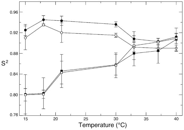
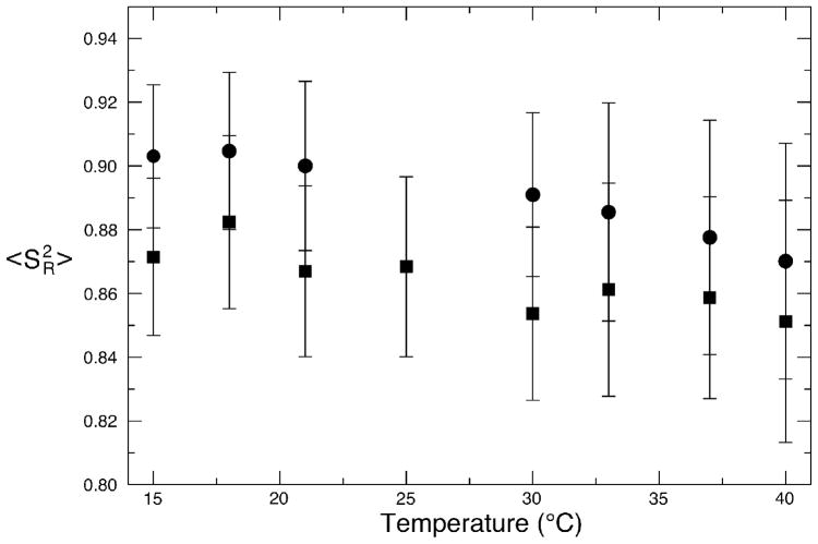
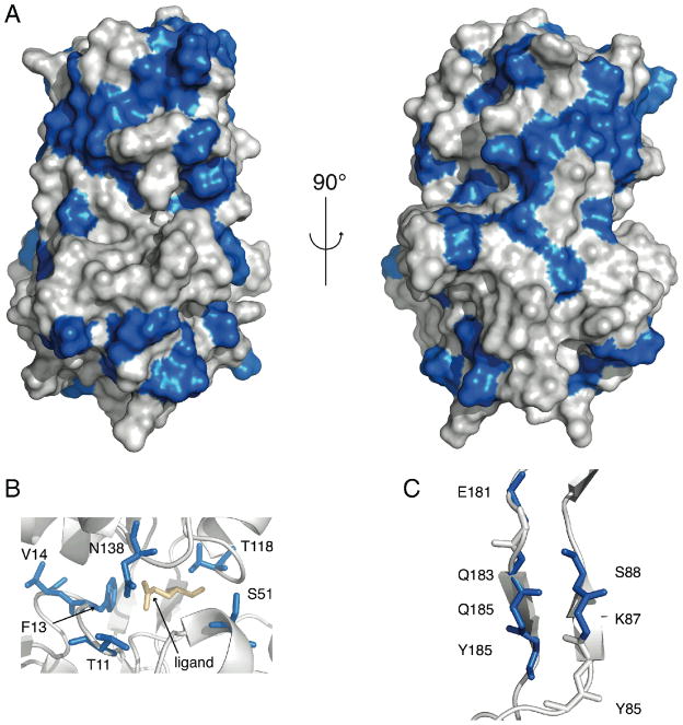
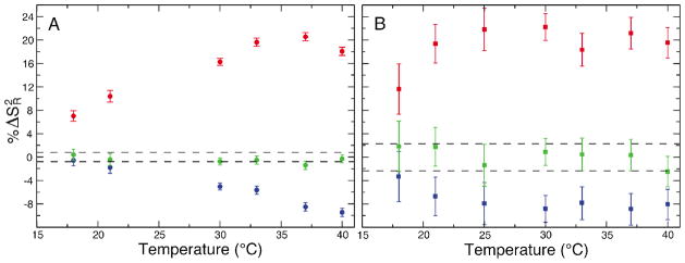
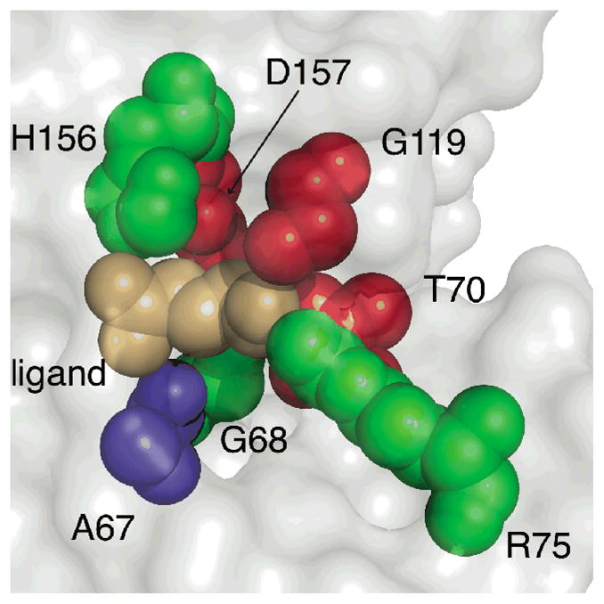
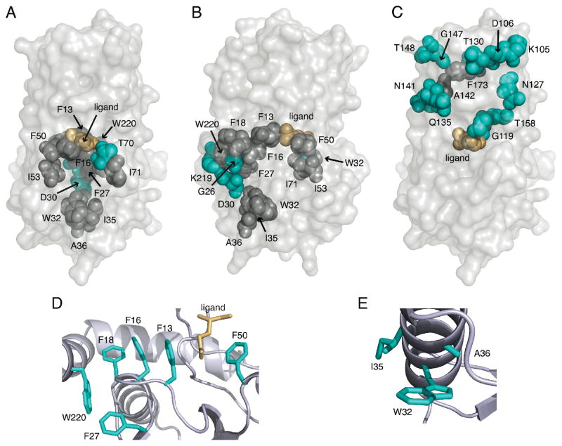
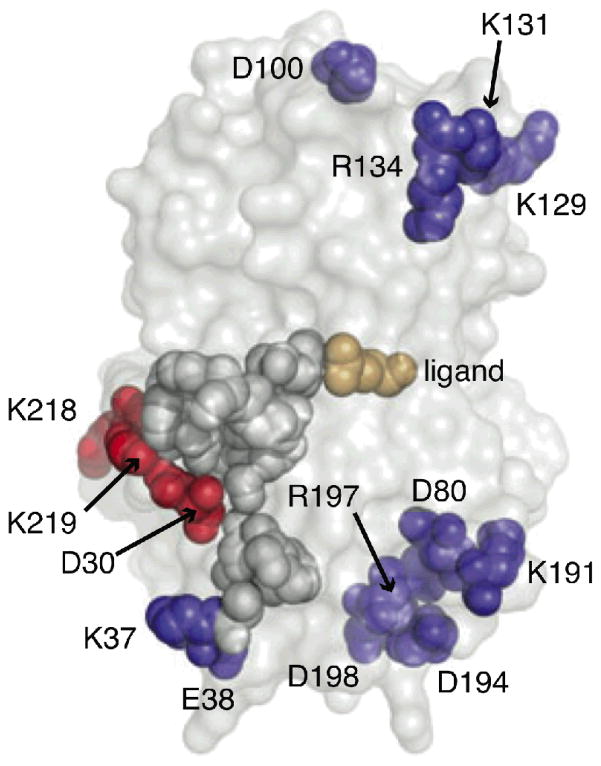
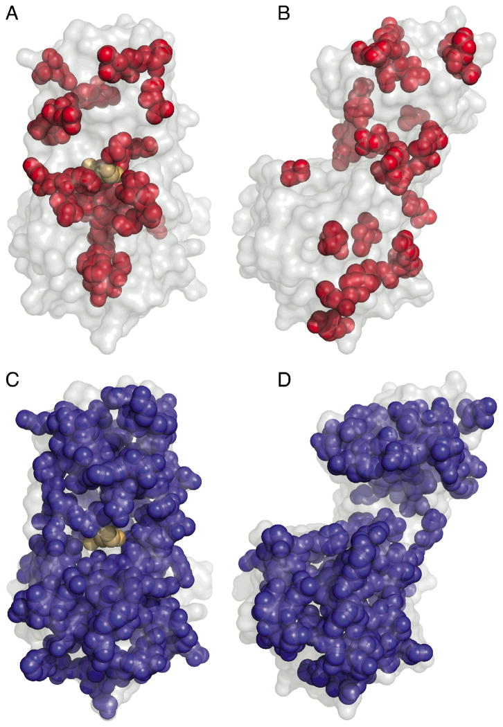
The temperature dependence of dynamic parameters derived from nuclear magnetic resonance (NMR) relaxation data is related to conformational entropy of the system under study. This provides information such as macromolecules stability and thermodynamics of ligand binding. We studied the temperature dependence of NMR order parameter of glutamine binding protein (GlnBP), a periplasmic binding protein (PBP) highly specific to L-glutamine associated with its ABC transporter, with the goal of elucidating the dynamical differences between the respective ligand bound and free forms. We found that the protein-ligand interaction, which is stabilized at higher temperature, has a striking effect on the stability of the hydrophobic core of the large domain of GlnBP. Moreover, in contrast to what was found for less specific PBPs, the decreasing backbone motion of the hinge region at increasing temperature supports the idea that the likelihood that GlnBP can adopt a ligand free closed conformation in solution diminishes at higher temperatures. Our results support the induced-fit model as mode of action for GlnBP. In addition, we found that the backbones of residues involved in a salt bridge do not necessarily become more rigid as the temperature rises as it was previously suggested [Vinther, J. M., et al. (2011) J. Am. Chem. Soc., 133, 271-278]. Our results show that for this to happen these residues have to also directly interact with a region of the protein that is becoming more rigid as the temperature increases.
Figures
Similar articles
-
Ligand-free open-closed transitions of periplasmic binding proteins: the case of glutamine-binding protein.Biochemistry. 2010 Mar 9;49(9):1893-902. doi: 10.1021/bi902045p. Biochemistry. 2010. PMID: 20141110 Free PMC article.
-
The structure of glutamine-binding protein complexed with glutamine at 1.94 A resolution: comparisons with other amino acid binding proteins.J Mol Biol. 1998 Apr 24;278(1):219-29. doi: 10.1006/jmbi.1998.1675. J Mol Biol. 1998. PMID: 9571045
-
Interdomain dynamics and ligand binding: molecular dynamics simulations of glutamine binding protein.FEBS Lett. 2003 Aug 28;550(1-3):168-74. doi: 10.1016/s0014-5793(03)00866-4. FEBS Lett. 2003. PMID: 12935905
-
The crystal structure of glutamine-binding protein from Escherichia coli.J Mol Biol. 1996 Sep 20;262(2):225-42. doi: 10.1006/jmbi.1996.0509. J Mol Biol. 1996. PMID: 8831790 Review.
-
Conformational dynamics and thermodynamics of protein-ligand binding studied by NMR relaxation.Biochem Soc Trans. 2012 Apr;40(2):419-23. doi: 10.1042/BST20110750. Biochem Soc Trans. 2012. PMID: 22435823 Review.
Cited by
-
Ligand-bound glutamine binding protein assumes multiple metastable binding sites with different binding affinities.Commun Biol. 2020 Aug 3;3(1):419. doi: 10.1038/s42003-020-01149-z. Commun Biol. 2020. PMID: 32747735 Free PMC article.
-
Dietary High Zinc Oxide Modulates the Microbiome of Ileum and Colon in Weaned Piglets.Front Microbiol. 2017 May 9;8:825. doi: 10.3389/fmicb.2017.00825. eCollection 2017. Front Microbiol. 2017. PMID: 28536569 Free PMC article.
-
NMR Analysis of Apo Glutamine-Binding Protein Exposes Challenges in the Study of Interdomain Dynamics.Angew Chem Int Ed Engl. 2019 Nov 18;58(47):16899-16902. doi: 10.1002/anie.201911015. Epub 2019 Oct 11. Angew Chem Int Ed Engl. 2019. PMID: 31515908 Free PMC article.
References
-
- Oswald C, Smits SHJ, Höing M, Sohn-Bösser L, Dupont L, Le Rudulier D, Schmitt L, Bremer E. Crystal Structures of the Choline/Acetylcholine Substrate-binding Protein ChoX from Sinorhizobium meliloti in the Liganded and Unliganded-Closed States. Journal of Biological Chemistry. 2008;283:32848–32859. - PubMed
-
- Tang C, Schwieters CD, Clore GM. Open-to-closed transition in apo maltose-binding protein observed by paramagnetic NMR. Nature. 2007;449:1078–1082. - PubMed
-
- Flocco MM, Mowbray SL. The 1.9 A x-ray structure of a closed unliganded form of the periplasmic glucose/galactose receptor from Salmonella typhimurium. Journal of Biological Chemistry. 1994;269:8931–8936. - PubMed
-
- Weiner JH, Heppel LA. A Binding Protein for Glutamine and Its Relation to Active Transport in Escherichia coli. Journal of Biological Chemistry. 1971;246:6933–6941. - PubMed
Publication types
MeSH terms
Substances
Grants and funding
LinkOut - more resources
Full Text Sources
Molecular Biology Databases
Research Materials
Miscellaneous
