Editorial
. 2011 Dec 29:9:90.
doi: 10.1186/1741-7007-9-90.

The bending of cell sheets--from folding to rolling

Affiliations
Editorial

The bending of cell sheets--from folding to rolling

Ray Keller et al. BMC Biol. .

Abstract

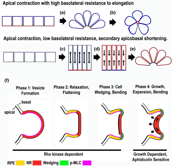
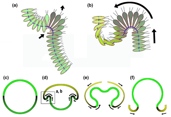
The bending of cell sheets plays a major role in multicellular embryonic morphogenesis. Recent advances are leading to a deeper understanding of how the biophysical properties and the force-producing behaviors of cells are regulated, and how these forces are integrated across cell sheets during bending. We review work that shows that the dynamic balance of apical versus basolateral cortical tension controls specific aspects of invagination of epithelial sheets, and recent evidence that tissue expansion by growth contributes to neural retinal invagination in a stem cell-derived, self-organizing system. Of special interest is the detailed analysis of the type B inversion in Volvox reported in BMC Biology by Höhn and Hallmann, as this is a system that promises to be particularly instructive in understanding morphogenesis of any monolayered spheroid system.


Figures

Figure 1
Figure 1
Sectional diagrams showing modes of epithelial bending. During monophasic epitheilial bending apical contraction occurs with relatively high basolateral resistance to cell elongation (a,b). In the biphasic mode, apical contraction occurs in the presence of relatively low resistance to elongation (c); this is followed by a second phase of apical-basal shortening in the presence of contracted apices, which results in bending (d,e) [2,3,5]. (f) Sectional diagrams illustrating bending of the neural retina epithelium in a stem cell-derived mammalian optic vesicle in culture [6]. RPE = retinal pigment epithelium; NR = neural retina; p-MLC = phosphorylated myosin light chain.
Figure 2
Figure 2
A sectional diagram illustrating aspects of the type B inversion in Volvox. (a,b) The initial steps in forming the subequatorial zone of cell wedging that progresses posteriorly and rolls or involutes (curved arrow) the posterior half into the anterior half are shown. (c-f) The location of this event in the context of the entire inversion is shown (box in (d)). The direction of the progression of zones of cell shape change is indicated by shading from dark to light (see text for discussion). Red, microtubules; blue, kinesins; black, cytoplasmic bridges. Based on [7-9].

References

    1. Hardin JD, Keller RE. The role of bottle cells in gastrulation of Xenopus. Development. 1988;103:210–230. - PubMed
    1. Keller RE, Davidson LA, Shook D. How we are shaped: The biomechanics of gastrulation. Differentiation. 2003;71:171–205. doi: 10.1046/j.1432-0436.2003.710301.x. - DOI - PubMed
    1. Sweeton D, Parks S, Costa M, Wieschaus E. Gastrulation in Drosophila: The formation of the ventral furrow and posterior midgut invaginations. Development. 1991;112:775–789. - PubMed
    1. Sherrard K, Robin F, Lemaire P, Munro E. Sequential activation of apical and basolateral contractility drives ascidian endoderm invagination. Curr Biol. 2010;20:1499–1510. doi: 10.1016/j.cub.2010.06.075. - DOI - PMC - PubMed
    1. Davidson LA, Keller R, Koehl M. Measurements of mechanical properties of the blastula wall reveal which hypothesized mechanisms of primary invagination are physically plausible in the sea urchin Strongylocentrotus purpuratus. Dev Biol. 1999;209:221–238. doi: 10.1006/dbio.1999.9249. - DOI - PubMed

Publication types

LinkOut - more resources