. 2011 Dec 29;11(1):44.
doi: 10.1186/1475-2867-11-44.

Multi-tyrosine kinase inhibitors in preclinical studies for pediatric CNS AT/RT: Evidence for synergy with Topoisomerase-I inhibition

Affiliations

Multi-tyrosine kinase inhibitors in preclinical studies for pediatric CNS AT/RT: Evidence for synergy with Topoisomerase-I inhibition

Aarthi Jayanthan et al. Cancer Cell Int. .

Abstract

Background: Currently, Atypical Teratoid Rhabdoid Tumor (AT/RT) constitutes one of the most difficult to treat malignancies in pediatrics. Hence, new knowledge of potential targets for therapeutics and the development of novel treatment approaches are urgently needed. We have evaluated the presence of cytokine pathways and the effects of two clinically available multi-tyrosine kinase inhibitors for cytotoxicity, target modulation and drug combinability against AT/RT cell lines.

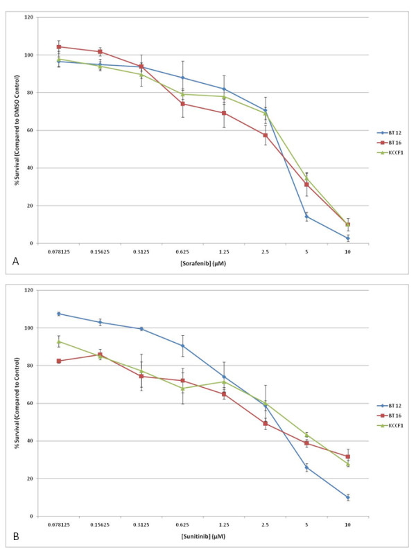
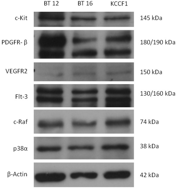
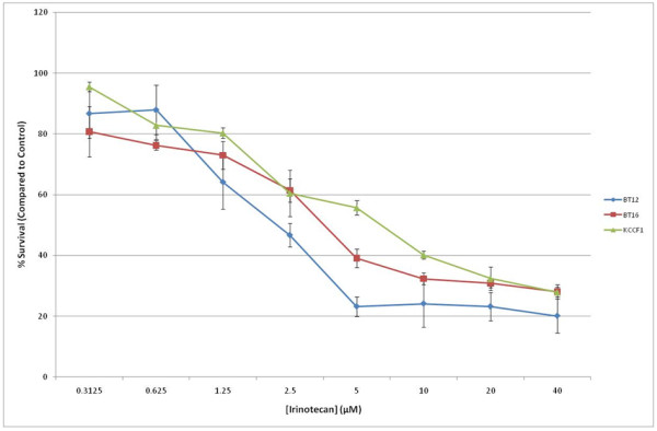
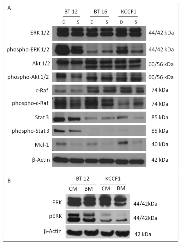
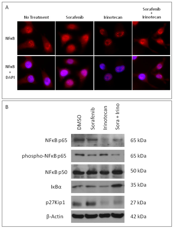
Results: AT/RT cell lines expressed measurable quantities of VEGF, FGF, PDGF and SDF-1, although the absolute amounts varied between the cell lines. The targeted receptor tyrosine kinase inhibitor sorafenib inhibited the key signaling molecule Erk, which was activated following the addition of own conditioned media, suggesting the existence of autocrine/paracrine growth stimulatory pathways. The multi-tyrosine kinase inhibitors sorafenib and sunitinib also showed significant growth inhibition of AT/RT cells and their activity was enhanced by combination with the topoisomerase inhibitor, irinotecan. The loss of cytoplasmic NF-kappa-B in response to irinotecan was diminished by sorafenib, providing evidence for a possible benefit for this drug combination.

Conclusions: In addition to previously described involvement of insulin like growth factor (IGF) family of cytokines, a multitude of other growth factors may contribute to the growth and survival of AT/RT cells. However, consistent with the heterogeneous nature of this tumor, quantitative and qualitative differences may exist among different tumor samples. Multi-tyrosine kinase inhibitors appear to have effective antitumor activity against all cell lines studied. In addition, the target modulation studies and drug combinability data provide the groundwork for additional studies and support the evaluation of these agents in future treatment protocols.


Figures

Figure 1
Figure 1
Sorafenib and sunitinib induce cell growth inhibition in AT/RT cell lines in vitro. Triplicate cultures of three AT/RT cell lines were incubated with increasing concentrations of sorafenib (A) or sunitinib (B). Appropriate dilutions of DMSO were also included as vehicle control. After four days in culture, cell viability was measured and plotted as a percentage of cell survival compared to equivalent vehicle control. Bars indicate SD. Data presented above are representative of five separate experiments. IC50 values calculated from these data are given in Table 2.
Figure 2
Figure 2
Expression of targets of sorafenib and sunitinib in AT/RT cell lines. Using Western blot analysis, it was determined that AT/RT cell lines express receptor tyrosine kinase targets of sorafenib and sunitinib: c-Kit, PDGF-Rβ, VEGFR2 and Flt-3. In addition, these cell lines express intracellular targets of sorafenib: c-Raf and p38α.
Figure 3
Figure 3
Irinotecan inhibits the in vitro growth of AT/RT cells. Triplicate cultures of AT/RT cell lines were cultured in the presence of increasing concentrations of irinotecan. Following four days in culture, cell growth inhibition was measured by Alamar blue assay. Percentage of cell survival was calculated by comparison of treated cells to control cells. Similar results were obtained in five separate experiments. IC50 values obtained from these data are given in Table 2.
Figure 4
Figure 4
The multi-kinase inhibitor sorafenib induces alterations in cellular signaling and apoptosis regulators in AT/RT cells in response to conditioned medium. Addition of autologous conditioned medium to briefly serum starved cells provides an avenue to evaluate the potential responses that may occur during autocrine/paracrine growth stimulation. In the presence of sorafenib (S), the conditioned medium induced reduced phosphorylation of Erk1/2 (BT12, KCCF1), Akt1/2 (BT12), c-Raf (BT12), Stat3 (BT12, KCCF1) compared to DMSO (D) control. Under these conditions, an increase in phosphorylation of c-Raf was noted in KCCF1 cells. However, the pro-survival protein Mcl-1 was suppressed by sorafenib in all three cell lines (A). Compared to basal medium (BM), conditioned medium (CM) induced phosphorylation of signaling regulators, such as Erk (B). The data shown are typical of three separate experiments.
Figure 5
Figure 5
Cytoplasmic NF-κB levels are lowered by irinotecan but sorafenib reduces this effect. BT12 cells were incubated with sorafenib or vehicle for 30 minutes followed by treatment with irinotecan for an additional 2 hours. For indirect immunofluorescence (A), cells were fixed and stained with antibodies to NF-κB followed by fluorescent labeled secondary antibodies. Concurrent DAPI stain was used to localize the nuclei (lower panel). Slides were visualized using a fluorescent microscope and random fields were photographed. The cytoplasmic staining seen in untreated and sorafenib treated cells was significantly reduced following treatment with irinotecan. However, the addition of sorafenib enabled the cells to maintain cytoplasmic staining in the presence of irinotecan. For Western blot analysis (B), cytoplasmic proteins were analyzed using antibodies against NFκB p65 and p50, phospho-NFκB p65, IκBα and p27Kip1. In the presence of irinotecan, there was a loss of cytoplasmic NF-κBp65, but in the presence of sorafenib, this loss was greatly reduced, corresponding to a decrease in phosphorylation of NF-κBp65. In addition, compared to treatment with sorafenib or irinotecan alone, there was increased expression of IκBα following treatment with sorafenib and irinotecan. Lastly, following treatment with irinotecan and sorafenib irinotecan combination, there was decreased expression of p27Kip1 compared to sorafenib treatment alone.

Similar articles

Cited by

References

    1. Bikowska B, Grajkowska W, Jozwiak J. Atypical teratoid/rhabdoid tumor: short clinical description and insight into possible mechanism of the disease. Eur J Neurol. 2011;18:813–818. doi: 10.1111/j.1468-1331.2010.03277.x. - DOI - PubMed
    1. Biegel JA. Molecular genetics of atypical teratoid/rhabdoid tumor. Neurosurg Focus. 2006;20:E11. doi: 10.3171/foc.2006.20.4.6. - DOI - PubMed
    1. Dufour C, Beaugrand A, Le Deley MC, Bourdeaut F, André N, Leblond P, Bertozzi AI, Frappaz D, Rialland X, Fouyssac F, Edan C, Grill J, Quidot M, Varlet P. Clinicopathologic prognostic factors in childhood atypical teratoid and rhabdoid tumor of the central nervous system: A multicenter study. Cancer. 2011. Electronically published ahead of print. - PubMed
    1. Bouvier C, De Paula AM, Fernandez C, Quilichini B, Scavarda D, Gentet JC, Figarella-Branger D. Atypical teratoid/rhabdoid tumor: 7-year event-free survival with gross total resection and radiotherapy in a 7-year-old boy. Childs Nerv Syst. 2008;24:143–147. - PubMed
    1. Guidi CJ, Sands AT, Zambrowicz BP, Turner TK, Demers DA, Webster W, Smith TW, Imbalzano AN, Jones SN. Disruption of Ini1 leads to peri-implantation lethality and tumorigenesis in mice. Mol Cell Biol. 2001;21:3598–3603. doi: 10.1128/MCB.21.10.3598-3603.2001. - DOI - PMC - PubMed