. 2012 Mar 1;363(1):40-51.
doi: 10.1016/j.ydbio.2011.12.021. Epub 2011 Dec 20.

Oocyte specific oolemmal SAS1B involved in sperm binding through intra-acrosomal SLLP1 during fertilization

Affiliations

Oocyte specific oolemmal SAS1B involved in sperm binding through intra-acrosomal SLLP1 during fertilization

Monika Sachdev et al. Dev Biol. .

Abstract

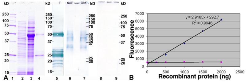
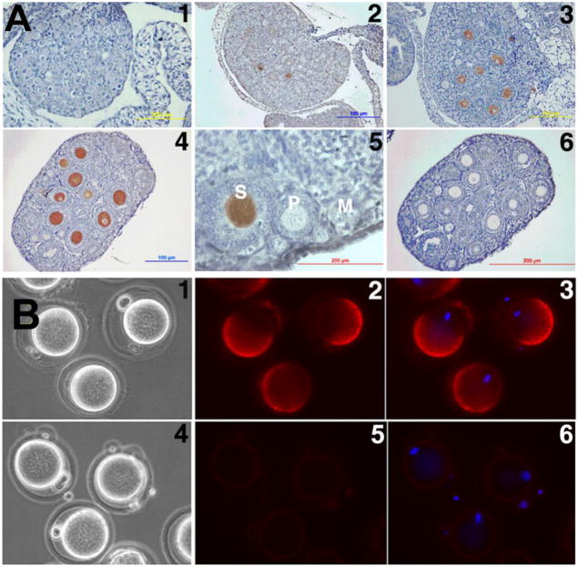
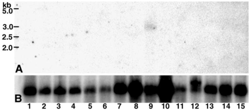
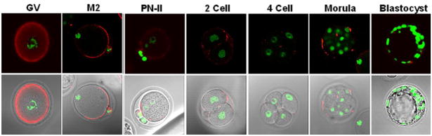
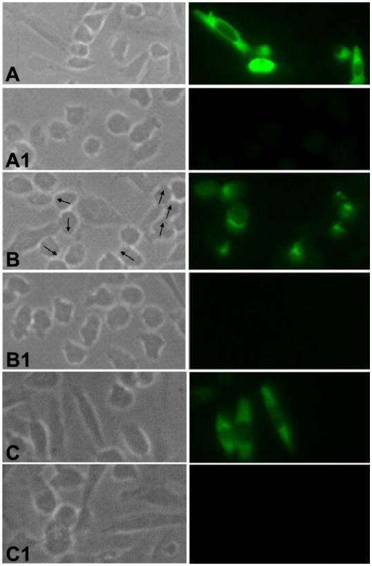
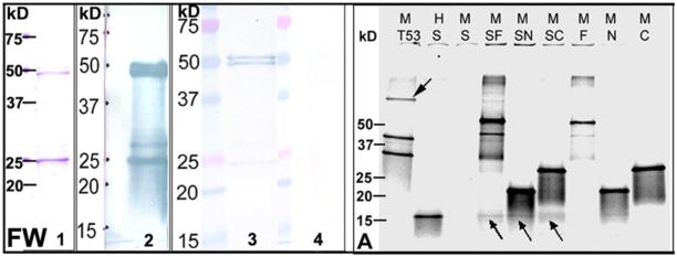
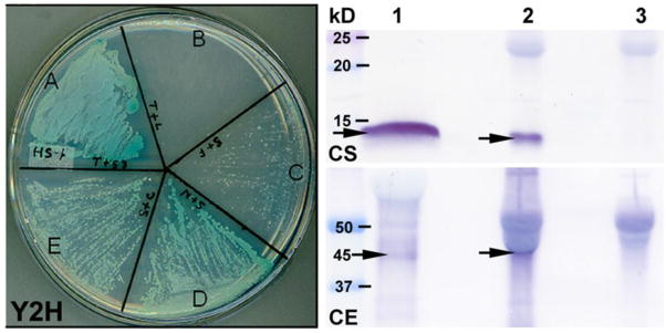
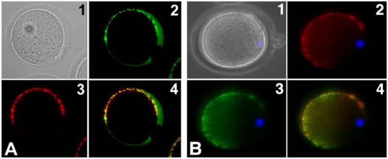
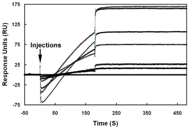
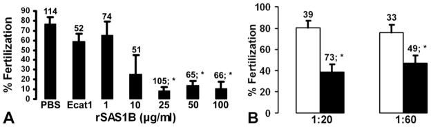
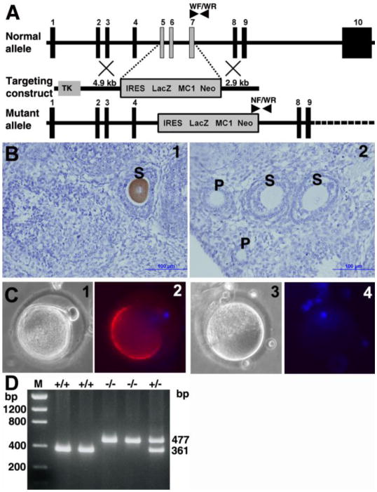
Molecular mechanisms by which fertilization competent acrosome-reacted sperm bind to the oolemma remain uncharacterized. To identify oolemmal binding partner(s) for sperm acrosomal ligands, affinity panning was performed with mouse oocyte lysates using sperm acrosomal protein, SLLP1 as a target. An oocyte specific membrane metalloproteinase, SAS1B (Sperm Acrosomal SLLP1 Binding), was identified as a SLLP1 binding partner. cDNA cloning revealed six SAS1B splice variants, each containing a zinc binding active site and a putative transmembrane domain, with signal peptides in three variants. SAS1B transcripts were ovary specific. SAS1B protein was first detected in early secondary follicles in day 3 ovaries. Immunofluorescence localized SAS1B to the microvillar oolemma of M2 oocytes. After fertilization, SAS1B decreased on the oolemma and became virtually undetectable in blastocysts. In transfected CHO-K1 cells SAS1B localized to the surface of unpermeabilized cells. Recombinant and native SLLP1 co-localized with SAS1B to the microvillar domain of ovulated M2 oocytes. Molecular interactions between mouse SLLP1 and SAS1B were demonstrated by surface plasmon resonance, far-western, yeast two-hybrid, recombinant- and native-co-IP analyses. SAS1B bound to SLLP1 with high affinity. SAS1B had protease activity, and SAS1B protein or antibody significantly inhibited fertilization. SAS1B knockout female mice showed a 34% reduction in fertility. The study identified SAS1B-SLLP1 as a pair of novel sperm-egg binding partners involving the oolemma and intra-acrosomal compartment during fertilization.


Figures

Figure 1
Figure 1
Expression, purification, Western and protease activity of rSAS1B. (A) The mature protein was expressed in E. coli, uninduced (L2), induced (L3), purified (L4, 3 μg) and stained with Coomassie, protein standards (L1). All purified bands were confirmed as rSAS1B by anti-His tag Western analysis (L5). SAS1B antibody recognized a range of native proteins from ~54 to ~31 kDa in zona intact (L6) and zona free (L7) oocyte protein extracts but no proteins were identified with preimmune antibody in the identical extracts (L8 and L9). (B) Assay of protease activity of rSAS1B (◆) using a fluorescent tagged synthetic peptide as substrate. Varying concentrations of purified proteins were used in 100 μL assay system. rSLLP1 (■) was used as a negative control.
Figure 2
Figure 2
Localization of SAS1B in developing ovary by IHC and in M2 oocytes by IF. (A) SAS1B expressed in ooplasm of secondary (S) follicles beginning in day 3 ovary (A2). No SAS1B was detected in naked oocytes (A1), primordial (M) and primary (P) follicles (A5). Panels 1, 2, 3, 4, 5 and 6 represent postpartum ovaries at day 0, 3, 7, 14, 56 and 14 respectively. Preimmune control, A6. (B) In unpermeablized zona-intact ovulated M2 oocytes, SAS1B was concentrated in a dome shaped microvillar domain on the surface of the oocyte plasma membrane. Eccentric nuclei (blue) were antipodal to the SAS1B positive domain. Preimmune control panels: 4, 5 and 6. Panels: 1 and 4, phase; 2 and 5, fluorescence; 3 and 6, merged image of DAPI/fluorescence.
Figure 3
Figure 3
Tissue expression of SAS1B transcript. A 15 mouse tissue Northern blot containing total RNA probed with 32P labelled SAS1B cDNA revealed a SAS1B mRNA of ~3.0 kb (A) only in ovary (lane 9). Same blot probed with GAPDH cDNA as control (B). Tissues: 1, brain; 2, stomach; 3, intestine; 4, colon; 5, liver; 6, lung; 7, kidney; 8, heart; 9, ovary; 10, muscle; 11, spleen; 12, testis; 13, thymus; 14, uterus; 15, placenta.
Figure 4
Figure 4
Confocal localization of SAS1B before and after fertilization. Oocytes and cultured early embryos stained with SAS1B antibody (red) and with Sytox (for nucleus, green, upper panel; merged on phase images, lower panel). SAS1B was localized throughout the cytoplasm of the GV oocyte where it concentrated at the cell periphery. After polar body formation, in M2 oocyte SAS1B localized mainly in the microvillar domain of oolemma. In fertilized oocyte (PN-II), SAS1B was located only in punctate regions at the cell periphery. These small patches persisted from 2-cell to morula stages when SAS1B appeared within the PVS. SAS1B was virtually undetectable in blastocyst stages.
Figure 5
Figure 5
SAS1B expression on the cell surface of transfected CHO-K1 cells. (A) In fixed and permeabilized cells SAS1B localized to cytoplasmic and surface domains. (B) Polarized localization (arrows) of SAS1B at the cell surface in fixed, unpermeabilized cells confirmed that SAS1B is a membrane protein. (C) Fixed and permeabilized cells probed with SAS1B C-terminal V5 tag antibody showing its presence in the cytoplasm. Panels: left, phase; right, fluorescence. The control cells (A1, B1, C1) that were transfected with the respective pcDNA3.1-TOPO vector lacking a SAS1B construct were probed with SAS1B specific antibodies revealing no immunofluorescence.
Figure 6
Figure 6
SAS1B-SLLP1 interactions by Far-Western (FW) and rCo-IP (A) analyses. (FW) Profile of purified rSAS1B stained with Coomassie used for FW analysis (L1). Western of rSAS1B probed with anti-his tag antibody (L2). rSAS1B blot either overlaid (L3) or not overlaid (L4) with purified soluble rSLLP1 (5 μg/ml) and probed with anti-SLLP1 monoclonal antibody. Full length SAS1B (~51, ~50 kD) bound to rSLLP1; however, the C-terminal ~25 kD protein band of similar intensity bound rSLLP1 only very weakly. (A) rCo-IP of SLLP1 using SAS1B myc-tag (M) antibody. Proteins were synthesized by in vitro translation in presence of S35-methionine and analyzed by SDS-PAGE. Translated SAS1B full length (F), N-terminus (N), C-terminus (C) and p53 (53) had myc-tag (M) while T-antigen (T) and SLLP1 (S) had HA-tag (H). Co-IP of T-antigen or SLLP1 (arrows) was done using anti-myc antibody from partner proteins. Co-IP reactions: T53, T-antigen + p53; SF, SLLP1 + SAS1B-F; SN, SLLP1 + SAS1B-N; SC, SLLP1 + SAS1B-C.
Fig. 7
Fig. 7
Y2H and native Co-IP analyses between SAS1B and SLLP1. Y2H assay showed affinity between SLLP1 and SAS1B fragments. Growth of yeast cells on high stringency plate forming blue colonies indicated interaction between the proteins. (A) Positive interaction between T-antigen and its partner p53. (B) Negative interaction between T-antigen and human lamin C. (C) Very weak interaction between full length SAS1B and SLLP1 with few small colonies but none visible at 3 days (Suppl. Fig. 6, HS). Positive interactions were noted between SLLP1 and N-terminal (D) or C-terminal forms (E). Native Co-IP of SAS1B from mixture of sperm and egg extracts using SLLP1 antibody. Equally divided eluted IP products were probed with SLLP1 (CS) or SAS1B antibody (CE). Sperm-egg extract IP by SLLP1 antibody revealed predominant ~45 kD SAS1B. L1, sperm or egg extract; L2, immune IP; L3, preimmune IP. Arrows indicate SLLP1 (~14 kD) or SAS1B (~45 kD).
Figure 8
Figure 8
Co-localization of recombinant SLLP1 and native SLLP1 with SAS1B. M2 oocytes incubated with rSLLP1 (A) or nSLLP1 (B), washed and probed with SLLP1 and SAS1B antibodies. SLLP1 (2) co-localized predominantly to the microvillar region of the mouse oolemma marked by SAS1B localization (3). Panels: 1, phase; 2, SLLP1; 3, SAS1B; 4, merge of 2 & 3; blue, nucleus.
Figure 9
Figure 9
Interaction affinity between SAS1B and SLLP1. Binding curves were observed when six different analyte concentrations of SAS1B (400 - top, 300, 200, 100, 50, 25 and 0 nM - bottom) were injected (arrow) for 3 min over immobilized SLLP1 at a flow rate of 30 μl/min in running buffer using Biacore 3000 system. Following the binding phase, the sensograms showed a very low dissociation of SAS1B from the chip.
Figure 10
Figure 10
SAS1B mediated inhibition of mouse in vitro fertilization. (A) Capacitated mouse sperm were incubated with varying concentrations of rSAS1B prior to fertilization of cumulus intact oocytes. The percentage of fertilized eggs decreased with increased concentration of SAS1B compared to a controls including the oocyte cytoplasmic protein recombinant Ecat1 or PBS without proteins. (B) Inhibition of fertilization of zona-intact eggs in presence of SAS1B antibody (black bar) compared to pre-immune (white bar) controls. Bars represent mean ± SEM; numbers, eggs per group. Significant P-value differences from no protein (PBS), recombinant Ecat1 (50 μg/ml) or preimmune controls were marked with asterisk (*, P ≤ 0.02 to 0.008).
Figure 11
Figure 11
Generation of SAS1B deficient mice. (A) Schematic representation of the targeting strategy of the Astl gene encoding SAS1B by homologous recombination. The targeting construct used thymidine kinase (TK) as a negative selection, MC1 Neo as a positive selection and IRES LacZ as a marker along with 4.9 kb and 2.9 kb homologous arms. The selection cassette targeted 3 coding exons (light vertical bars) 5, 6 and 7 encoding 131 residues (SPF – ILP) of the normal allele. Genotyping primer positions in normal and mutant alleles were indicated by arrow heads. (B) Immunohistochemical localization of SAS1B in wild type (1) and null (2) mice ovaries using anti-SAS1B guinea pig antibody confirmed the loss of SAS1B protein in oocytes within growing follicles of knockout animals. S, secondary follicles; P, primary follicles. (C) Immuno-fluorescence localization of SAS1B in wild type (1 and 2) and knockout (3 and 4) ovulated eggs showed lack of SAS1B in the microvillar domain of mature M2 egg in null mice. Panels: 1 and 3 phase; 2 and 4 merge image of DAPI/fluorescence. (D) Genotype PCR of genomic tail DNA using 3 primers (WF, WR, NF) producing a 361 bp product in wild type (+/+), a 477 bp product in null (−/−) and both products in heterozygous mice. M, DNA mass ladder.

References

    1. Alfieri JA, Martin AD, Takeda J, Kondoh G, Myles DG, Primakoff P. Infertility in female mice with an oocyte-specific knockout of GPI-anchored proteins. J Cell Sci. 2003;116:2149–2155. - PubMed
    1. Almeida EA, Huovila AP, Sutherland AE, Stephens LE, Calarco PG, Shaw LM, Mercurio AM, Sonnenberg A, Primakoff P, Myles DG. Mouse egg integrin α6β1 functions as a sperm receptor. Cell. 1995;81:1095–1104. - PubMed
    1. Cho C, Bunch DO, Faure JE, Goulding EH, Eddy EM, Primakoff P, Myles DG. Fertilization defects in sperm from mice lacking fertilin β. Science. 1998;281:1857–1859. - PubMed
    1. Cohen DJ, Ellerman DA, Cuasnicu PS. Mammalian sperm-egg fusion: evidence that epididymal protein DE plays a role in mouse gamete fusion. Biol Reprod. 2000;63:462–468. - PubMed
    1. Coonrod SA, Naaby-Hansen S, Shetty J, Shibahara H, Chen M, White JM, Herr JC. Treatment of mouse oocytes with PI-PLC releases 70-kDa (pI 5) and 35- to 45-kDa (pI 5.5) protein clusters from the egg surface and inhibits sperm-oolemma binding and fusion. Dev Biol. 1999;207:334–349. - PubMed

Publication types

MeSH terms