. 2012 Feb 17;416(2):208-20.
doi: 10.1016/j.jmb.2011.12.030. Epub 2011 Dec 21.

Molecular structure and peptidoglycan recognition of Mycobacterium tuberculosis ArfA (Rv0899)

Affiliations

Molecular structure and peptidoglycan recognition of Mycobacterium tuberculosis ArfA (Rv0899)

Yong Yao et al. J Mol Biol. .

Abstract

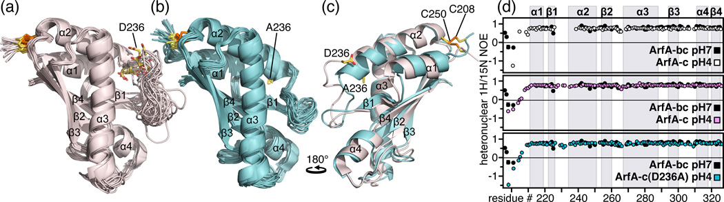
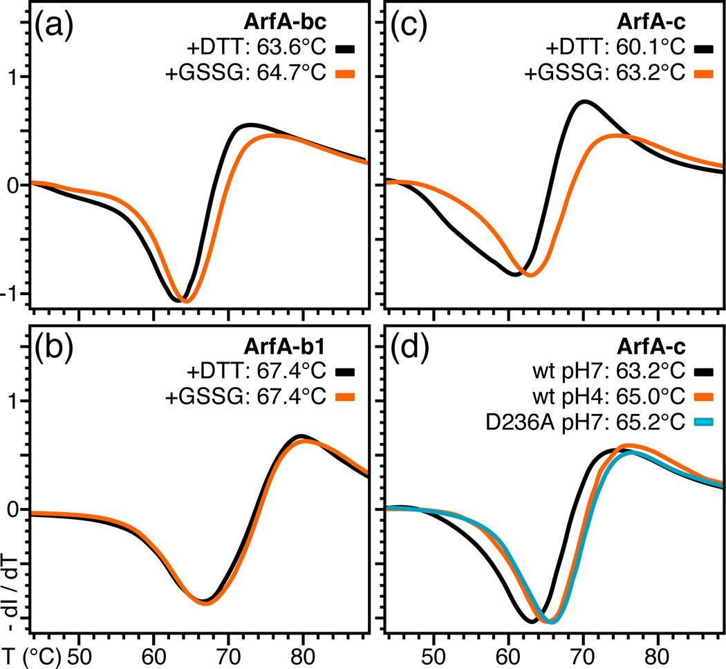
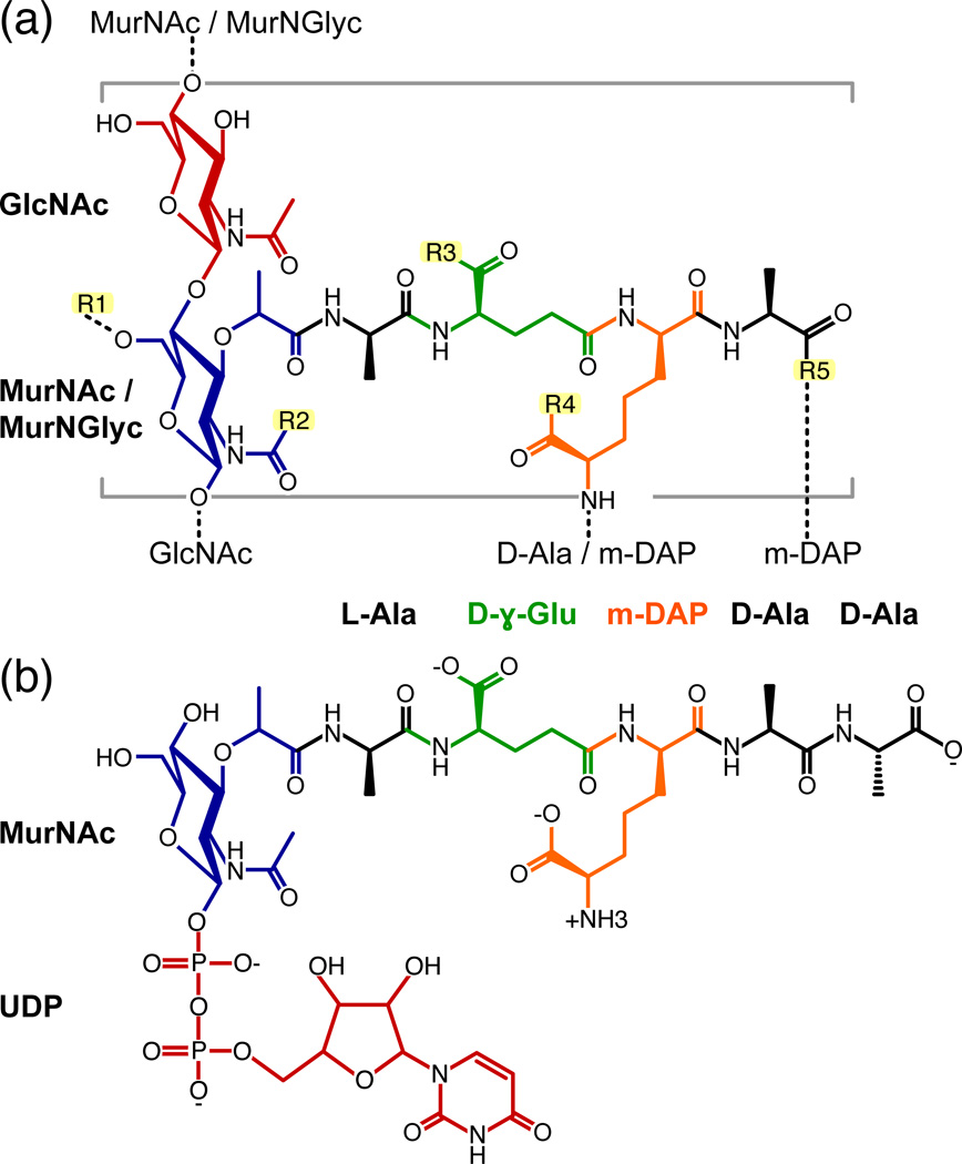
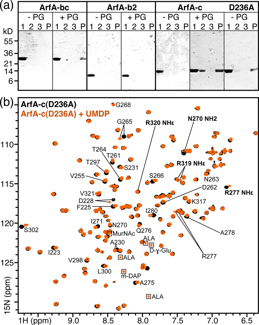
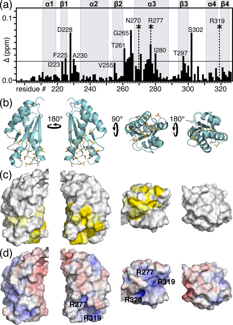
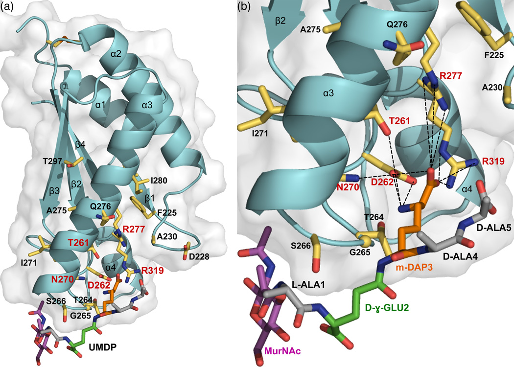
Mycobacterium tuberculosis ArfA (Rv0899) is a membrane protein encoded by an operon that is required for supporting bacterial growth in acidic environments. Its C-terminal domain (C domain) shares significant sequence homology with the OmpA-like family of peptidoglycan-binding domains, suggesting that its physiological function in acid stress protection may be related to its interaction with the mycobacterial cell wall. Previously, we showed that ArfA forms three independently structured modules, and we reported the structure of its central domain (B domain). Here, we describe the high-resolution structure and dynamics of the C domain, we identify ArfA as a peptidoglycan-binding protein and we elucidate the molecular basis for its specific recognition of diaminopimelate-type peptidoglycan. The C domain of ArfA adopts the characteristic fold of the OmpA-like family. It exhibits pH-dependent conformational dynamics (with significant heterogeneity at neutral pH and a more ordered structure at acidic pH), which could be related to its acid stress response. The C domain associates tightly with polymeric peptidoglycan isolated from M. tuberculosis and also associates with a soluble peptide intermediate of peptidoglycan biosynthesis. This enabled us to characterize the peptidoglycan binding site where five highly conserved ArfA residues, including two key arginines, establish the specificity for diaminopimelate- but not Lys-type peptidoglycan. ArfA is the first peptidoglycan-binding protein to be identified in M. tuberculosis. Its functions in acid stress protection and peptidoglycan binding suggest a link between the acid stress response and the physicochemical properties of the mycobacterial cell wall.


Figures

Figure 1
Figure 1. Structures and backbone dynamics of wild-type ArfA-c and ArfA-c(D236A)
(a, b) Backbone representations of the 20 lowest energy structures of (a) wild-type ArfA-c and (b) ArfA-c(D236A). (c) Superimposed lowest energy structures of ArfA-c (pink) and ArfA-c(D236A) (cyan). The structures were determined at pH7. The average pairwise RMSDs for the structured core of the protein (residues 207–326) are reported in Table S1. (d) Heteronuclear 1H/15N NOEs reflecting backbone dynamics of: ArfA-bc at pH7 (black), ArfA-c at pH7 (white), ArfA-c at pH4 (pink), and ArfA-c(D236A) at pH7 (cyan). For wild-type ArfA-c, peaks from residues in the β1-α2 loop could not be observed at pH7. Residues in this loop gave weak, broad peaks at pH4, but gave peaks of normal intensity in the D236A mutant.
Figure 2
Figure 2. Differential scanning fluorimetry traces showing the effect of the disulfide bond, pH, and D236A mutation on the thermal stability of ArfA
(a–c) Protein was treated with DTT or GSSG to break or form the C208–C250 disulfide bond. ArfA-b1 encompasses residues 73 to 220. (d) Traces were obtained for wild-type ArfA-c at pH7 (black) or pH4 (orange) and for ArfA-c(D236A) at pH7 (cyan).
Figure 3
Figure 3. Chemical structures of (a) M. tuberculosis peptidoglycan and (b) the peptidoglycan biosynthesis intermediate, UMDP
Dashed lines designate cross links to arabinogalactan or to the sugar and peptide stems of neighboring monomeric units. GlcNAc: N-acetyl-glucosamine; MurNGlyc: N-glycolyl-muramic acid; MurNAc: N-acetyl-muramic acid; m-DAP: meso-diaminopimelic acid. UDP: uridine-5'-diphosphate; R1: H or the linker unit of arabinogalactan; R2: H, CH3 (N-acetyl) or CH2OH (N-glycolyl); R3, R4: OH, NH2 or OCH3; R5: OH or amidated cross link to m-DAP.
Figure 4
Figure 4. Association of ArfA-c with peptidoglycan
(a) Soluble, purified ArfA polypeptides: ArfA-bc, ArfA-b2 (residues 73–197), ArfA-c and ArfA-c(D236A) were mixed with purified peptidoglycan from M. tuberculosis (+PG). As negative controls, proteins were incubated without peptidoglycan (−PG). After 2 hr incubation, the supernatant (Lane 1) and insoluble fraction were separated by centrifugation, the pellet was washed with buffer and again separated from the supernatant (Lane 2) by centrifugation. After a second wash and centrifugation step to generate a third supernatant (Lane 3) and final pellet (Lane P), all fractions were analyzed by SDS-PAGE and visualized with Coomassie blue. ArfA-bc, ArfA-c and ArfA-c(D236A) bind peptidoglycan and separate with it in the insoluble fraction, while ArfA-b2 does not bind peptidoglycan and remains in solution. For each ArfA polypeptide, the result of one out of two or more independent experiments is shown. (b) NMR 1H/15N HSQC spectra of ArfA-c(D236A) obtained at pH7, 25°C, with (orange) or without (black) ~20 molar equivalents of UMDP. Examples of peaks sensitive (e.g. D228) or insensitive (e.g. G268) to addition of UMDP are labeled. Peaks from side chains are labeled in bold. Peaks from the natural abundance peptide are enclosed in boxes.
Figure 5
Figure 5. Peptidoglycan binding site of M. tuberculosis ArfA-c(D236A)
(a) Chemical shift changes in the spectrum of ArfA-c(D236A) induced by UMDP. The total chemical shift change (Δ) for each peak was calculated by adding the changes in 1H (ΔH) and 15N (ΔN) using the equation Δ = [(ΔH)2 + (ΔN/5)2]1/2. Asterisks indicate side chain NH peaks that undergo significant chemical shift or intensity changes. Values of Δ≥0.03 ppm (horizontal line) were mapped on the structure. (b, c) Molecular backbone and surface representations of ArfA-c(D236A) showing residues with Δ≥0.03 ppm (yellow) in the peptidoglycan binding site of ArfA. (d) Molecular surface representations colored by electrostatic potential, with isocontours shown at +8kBT (blue) and −8kBT (red), where kB is the Boltzmann constant, and T is the temperature.
Figure 6
Figure 6. Peptidoglycan recognition by ArfA-c
(a) Full and (b) close-up views of the structure of ArfA-c(D236A) associated with a structural model of the peptidoglycan peptide MDP (MurNAc–L-Ala–D-γ-Glu–m-DAP–D-Ala–D-Ala). Residues with NMR peaks most affected by UMDP (Δ≥0.03 ppm) are shown as yellow sticks. Residues that form the m-DAP recognition site are labeled in red. MDP is color coded with MurNAc in magenta, Ala in gray, D-γ-Glu in green, and m-DAP in orange.

References

    1. Hett EC, Rubin EJ. Bacterial growth and cell division: a mycobacterial perspective. Microbiol Mol Biol Rev. 2008;72:126–156. - PMC - PubMed
    1. Kaur D, Guerin ME, Skovierova H, Brennan PJ, Jackson M. Chapter 2: Biogenesis of the cell wall and other glycoconjugates of Mycobacterium tuberculosis. Advances in Applied Microbiology. 2009;69:23–78. - PMC - PubMed
    1. Hoffmann C, Leis A, Niederweis M, Plitzko JM, Engelhardt H. Disclosure of the mycobacterial outer membrane: cryo-electron tomography and vitreous sections reveal the lipid bilayer structure. Proceedings of the National Academy of Sciences of the United States of America. 2008;105:3963–3967. - PMC - PubMed
    1. Zuber B, Chami M, Houssin C, Dubochet J, Griffiths G, Daffe M. Direct visualization of the outer membrane of mycobacteria and corynebacteria in their native state. J Bacteriol. 2008;190:5672–5680. - PMC - PubMed
    1. Sani M, Houben EN, Geurtsen J, Pierson J, de Punder K, van Zon M, Wever B, Piersma SR, Jimenez CR, Daffe M, Appelmelk BJ, Bitter W, van der Wel N, Peters PJ. Direct visualization by cryo-EM of the mycobacterial capsular layer: a labile structure containing ESX-1-secreted proteins. PLoS Pathog. 2010;6 e1000794. - PMC - PubMed

Publication types

MeSH terms

LinkOut - more resources