

Feasibility of sentinel node biopsy in head and neck melanoma using a hybrid radioactive and fluorescent tracer
- PMID: 22207047
- PMCID: PMC3356513
- DOI: 10.1245/s10434-011-2180-7
Feasibility of sentinel node biopsy in head and neck melanoma using a hybrid radioactive and fluorescent tracer
Abstract
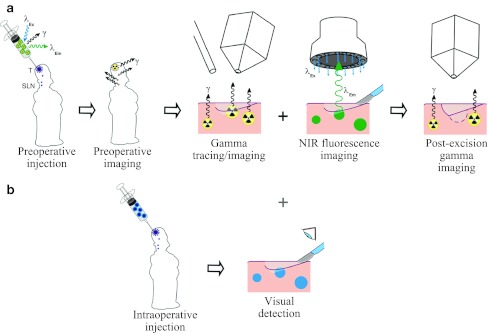
Purpose: This study was designed to examine the feasibility of combining lymphoscintigraphy and intraoperative sentinel node identification in patients with head and neck melanoma by using a hybrid protein colloid that is both radioactive and fluorescent.
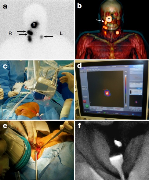
Methods: Eleven patients scheduled for sentinel node biopsy in the head and neck region were studied. Approximately 5 h before surgery, the hybrid nanocolloid labeled with indocyanine green (ICG) and technetium-99m ((99m)Tc) was injected intradermally in four deposits around the scar of the primary melanoma excision. Subsequent lymphoscintigraphy and single photon emission computed tomography with computed tomography (SPECT/CT) were performed to identify the sentinel nodes preoperatively. In the operating room, patent blue dye was injected in 7 of the 11 patients. Intraoperatively, sentinel nodes were acoustically localized with a gamma ray detection probe and visualized by using patent blue dye and/or fluorescence-based tracing with a dedicated near-infrared light camera. A portable gamma camera was used before and after sentinel node excision to confirm excision of all sentinel nodes.
Results: A total of 27 sentinel nodes were preoperatively identified on the lymphoscintigraphy and SPECT/CT images. All sentinel nodes could be localized intraoperatively. In the seven patients in whom blue dye was used, 43% of the sentinel nodes stained blue, whereas all were fluorescent. The portable gamma camera identified additional sentinel nodes in two patients. Ex vivo, all radioactive lymph nodes were fluorescent and vice versa, indicating the stability of the hybrid tracer.
Conclusions: ICG-(99m)Tc-nanocolloid allows for preoperative sentinel node visualization and concomitant intraoperative radio- and fluorescence guidance to the same sentinel nodes in head and neck melanoma patients.
Conflict of interest statement
The authors declare that they have no conflict of interest.
Figures
References
-
- Eicher SA, Clayman GL, Myers JN, Gillenwater AM. A prospective study of intraoperative lymphatic mapping for head and neck cutaneous melanoma. Arch Otolaryngol Head Neck Surg. 2002;128:241–246. - PubMed
Publication types
MeSH terms
Substances
LinkOut - more resources
Full Text Sources
Medical
