

18α-Glycyrrhizin induces apoptosis and suppresses activation of rat hepatic stellate cells
- PMID: 22207106
- PMCID: PMC3560665
- DOI: 10.12659/msm.882196
18α-Glycyrrhizin induces apoptosis and suppresses activation of rat hepatic stellate cells
Abstract
Background: To investigate the potential mechanisms underlying the protective effects of 18α Glycyrrhizin (GL) on rat hepatic stellate cells (HSCs) and hepatocytes in vivo and in vitro.
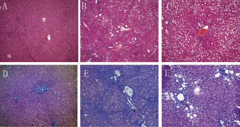
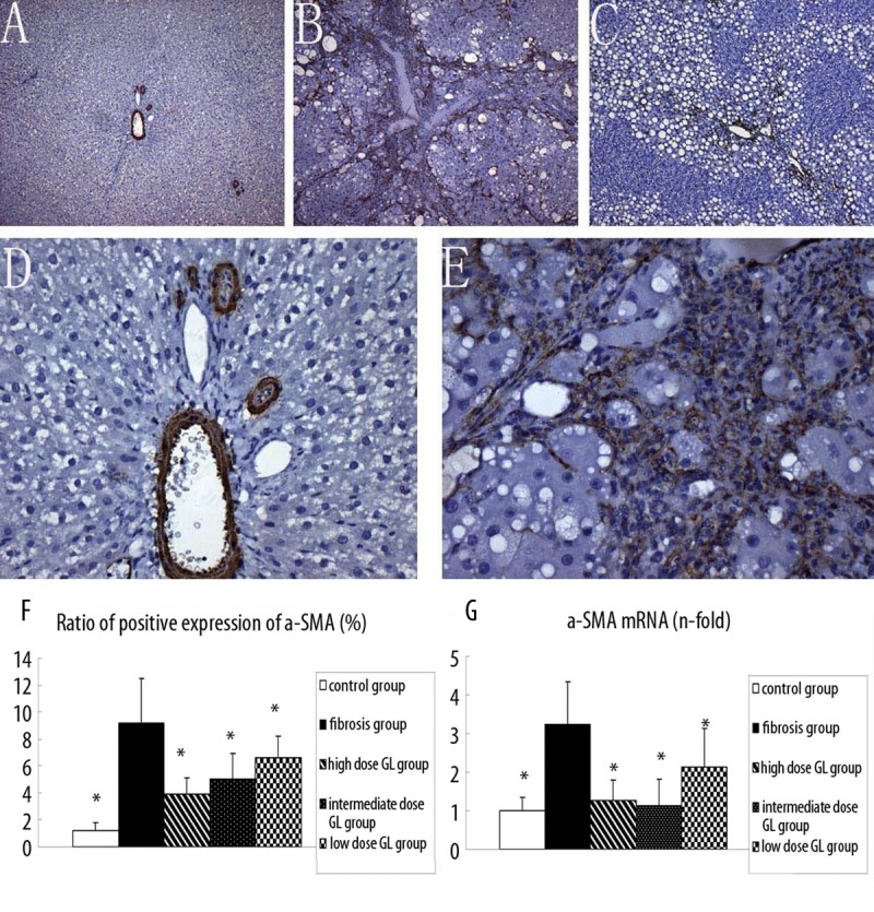
Material/methods: Sprague-Dawley (SD) rats were randomly divided into 5 groups: normal control group, liver fibrosis group, high-dose 18α GL group (25 mg/ kg/d), intermediate-dose 18α GL group (12.5 mg/kg/d) and low-dose 18α GL group (6.25 mg/ kg/d). The rat liver fibrosis model was induced by carbon tetrachloride (CCl4). The expressions of alpha-smooth muscle actin (αSMA) and NF-kappaB were determined by real-time PCR and immunohistochemistry.
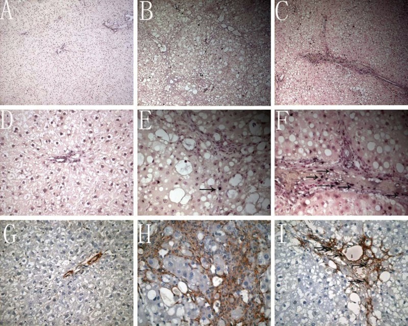
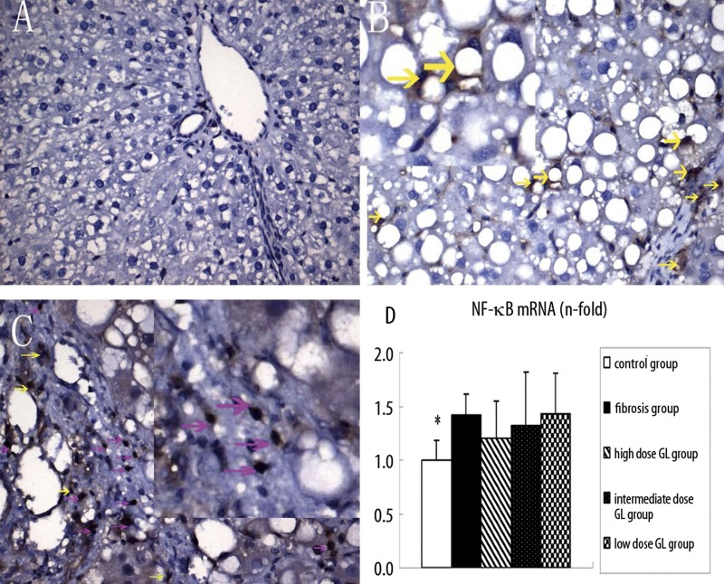
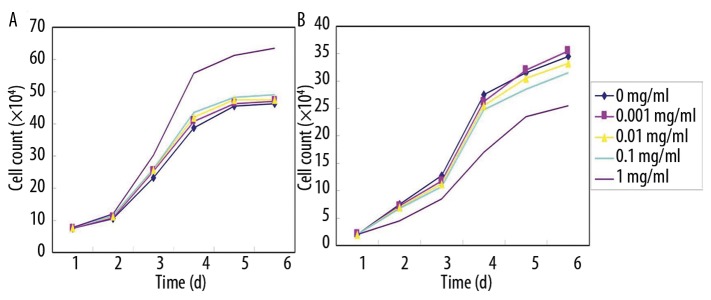
Results: 18αGL dose-dependently inhibited the CCl4-induced liver fibrosis. There were significant differences in the mRNA and protein expressions of αSMA between the fibrosis group and 18α-GL treatment groups, suggesting that 18α GL can suppress the proliferation and activation of HSCs. Few HSCs were apoptotic in the portal area and fibrous septum in the liver fibrosis group. However, the double-color staining of a-SMA and TUNEL showed that 18α-GL treatment groups increased HSC apoptosis. NF-kappaB was mainly found in the nucleus in the fibrosis group, while cytoplasmic expression of NF-kappaB was noted in the 18αGL groups. In the in vitro experiments, 18α GL promoted the proliferation of hepatocytes, but inhibited that of HSCs. HSCs were arrested in the G2/M phase following 18α GL treatment and were largely apoptotic.
Conclusions: 18α-GL can suppress the activation of HSCs and induce the apoptosis of HSCs by blocking the translocation of NF-kappaB into the nucleus, which plays an important role in the protective effect of 18α-GL on liver fibrosis.
Figures
Similar articles
-
Effects of 18α-glycyrrhizin on TGF-β1/Smad signaling pathway in rats with carbon tetrachloride-induced liver fibrosis.Int J Clin Exp Pathol. 2015 Feb 1;8(2):1292-301. eCollection 2015. Int J Clin Exp Pathol. 2015. PMID: 25973013 Free PMC article.
-
Effects of interleukin-10 on activation and apoptosis of hepatic stellate cells in fibrotic rat liver.World J Gastroenterol. 2006 Mar 28;12(12):1918-23. doi: 10.3748/wjg.v12.i12.1918. World J Gastroenterol. 2006. PMID: 16609999 Free PMC article.
-
The Salvia miltiorrhiza monomer IH764-3 induces apoptosis of hepatic stellate cells in vivo in a bile duct ligation-induced model of liver fibrosis.Mol Med Rep. 2012 Dec;6(6):1231-8. doi: 10.3892/mmr.2012.1076. Epub 2012 Sep 11. Mol Med Rep. 2012. PMID: 22971838
-
Meta-Analysis Based on Clinical RCTs: The Effect of Molecular Epimerism on the Safety of Glycyrrhizic Acid.Evid Based Complement Alternat Med. 2020 Nov 30;2020:3869698. doi: 10.1155/2020/3869698. eCollection 2020. Evid Based Complement Alternat Med. 2020. PMID: 33312222 Free PMC article. Review.
-
The role of p53 in liver fibrosis.Front Pharmacol. 2022 Oct 24;13:1057829. doi: 10.3389/fphar.2022.1057829. eCollection 2022. Front Pharmacol. 2022. PMID: 36353498 Free PMC article. Review.
Cited by
-
Efficacy and safety of glycyrrhizic acid preparation treating comorbid liver injury in COVID-19: A systematic review.Front Pharmacol. 2022 Nov 3;13:1003697. doi: 10.3389/fphar.2022.1003697. eCollection 2022. Front Pharmacol. 2022. PMID: 36408213 Free PMC article.
-
Synthesis and discovery of 18α-GAMG as anticancer agent in vitro and in vivo via down expression of protein p65.Sci Rep. 2014 Nov 19;4:7106. doi: 10.1038/srep07106. Sci Rep. 2014. PMID: 25407586 Free PMC article.
-
Liver fibrosis and protection mechanisms action of medicinal plants targeting apoptosis of hepatocytes and hepatic stellate cells.Adv Pharmacol Sci. 2014;2014:373295. doi: 10.1155/2014/373295. Epub 2014 Nov 20. Adv Pharmacol Sci. 2014. PMID: 25505905 Free PMC article. Review.
-
Protective mechanisms of medicinal plants targeting hepatic stellate cell activation and extracellular matrix deposition in liver fibrosis.Chin Med. 2014 Dec 24;9(1):27. doi: 10.1186/s13020-014-0027-4. eCollection 2014. Chin Med. 2014. PMID: 25606051 Free PMC article.
-
Synthesis, molecular docking and biological evaluation of glycyrrhizin analogs as anticancer agents targeting EGFR.Molecules. 2014 May 19;19(5):6368-81. doi: 10.3390/molecules19056368. Molecules. 2014. PMID: 24853453 Free PMC article.
References
-
- Friedman SL. Liver fibrosis – from bench to bedside. J Hepatol. 2003;38:S38–53. - PubMed
-
- Gressner AM. The cell biology of liver fibrogenesis – an imbalance of proliferation, growth arrest and apoptosis of myofibroblasts. Cell Tissue Res. 1998;292:447–52. - PubMed
-
- Eisenbrand G. Glycyrrhizin. Mol Nutr Food Res. 2006;50:1087–88. - PubMed
-
- Wu X, Zhang L, Gurley E, et al. Prevention of free fatty acid-induced hepatic lipotoxicity by 18beta-glycyrrhetinic acid through lysosomal and mitochondrial pathways. Hepatology. 2008;47(6):1905–15. - PubMed
-
- Scheuer PJ. Classification of chronic viral hepatitis: a need for reassessment. J Hepatol. 1991;13:372–74. - PubMed
Publication types
MeSH terms
Substances
LinkOut - more resources
Full Text Sources
Other Literature Sources
Medical
