

A tolerogenic role for Toll-like receptor 9 is revealed by B-cell interaction with DNA complexes expressed on apoptotic cells
- PMID: 22207622
- PMCID: PMC3271931
- DOI: 10.1073/pnas.1109173109
A tolerogenic role for Toll-like receptor 9 is revealed by B-cell interaction with DNA complexes expressed on apoptotic cells
Abstract
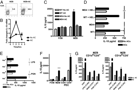
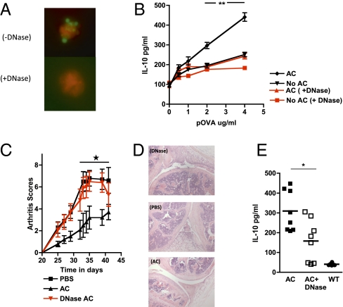
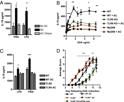
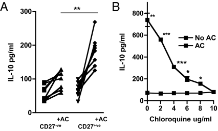
Intracellular protein complexes containing nucleic acids are common targets of autoantibodies in many autoimmune diseases. Central tolerance to these antigens is incomplete, yet nucleosomal DNA is expressed on the surface of cells dying by apoptosis. It is commonly believed that autoimmunity is prevented by the rapid uptake of apoptotic cells (ACs) by neighbors or professional phagocytes to which they deliver anti-inflammatory signals. Self-reactive, innate-like B cells contact and are selected by intracellular antigens expressed on ACs; however, how self-tolerance is maintained is not well understood. Here we report that IL-10 production by B cells, stimulated by contact with ACs, results from the engagement of Toll-like receptor 9 (TLR9) within the B cell after recognition of DNA-containing complexes on the surface of ACs. Until now, TLR9 ligation has been considered an inflammatory signal, but we have confirmed a hitherto unexpected immunoregulatory role by demonstrating the absence of the protective effect of ACs during experimental autoimmune encephalitis (EAE) in TLR9-deficient mice. Human circulating CD27(+) B cells also respond to DNA-bearing ACs, but not to DNase-treated cells, by secreting IL-10. Chronic autoimmune disease may arise if this tolerance mechanism is not reimposed after episodes of inflammation, or if the regulatory B-cell response is subverted.
Conflict of interest statement
The authors declare no conflict of interest.
Figures
Similar articles
-
Signaling via toll-like receptor 4 and CD40 in B cells plays a regulatory role in the pathogenesis of multiple sclerosis through interleukin-10 production.J Autoimmun. 2018 Mar;88:103-113. doi: 10.1016/j.jaut.2017.10.011. Epub 2017 Nov 13. J Autoimmun. 2018. PMID: 29146546
-
Regulatory B cells mediate tolerance to apoptotic self in health: implications for disease.Int Immunol. 2015 Oct;27(10):505-11. doi: 10.1093/intimm/dxv045. Epub 2015 Aug 25. Int Immunol. 2015. PMID: 26306497 Review.
-
A TLR9-dependent checkpoint governs B cell responses to DNA-containing antigens.J Clin Invest. 2017 May 1;127(5):1651-1663. doi: 10.1172/JCI89931. Epub 2017 Mar 27. J Clin Invest. 2017. PMID: 28346226 Free PMC article.
-
TLR9 in peritoneal B-1b cells is essential for production of protective self-reactive IgM to control Th17 cells and severe autoimmunity.J Immunol. 2011 Sep 15;187(6):2953-65. doi: 10.4049/jimmunol.1003340. Epub 2011 Aug 22. J Immunol. 2011. PMID: 21859955
-
Self-DNA at the Epicenter of SLE: Immunogenic Forms, Regulation, and Effects.Front Immunol. 2019 Jul 10;10:1601. doi: 10.3389/fimmu.2019.01601. eCollection 2019. Front Immunol. 2019. PMID: 31354738 Free PMC article. Review.
Cited by
-
Immunosuppressive Mechanisms of Regulatory B Cells.Front Immunol. 2021 Apr 29;12:611795. doi: 10.3389/fimmu.2021.611795. eCollection 2021. Front Immunol. 2021. PMID: 33995344 Free PMC article. Review.
-
Regulation of B Cell Responses in SLE by Three Classes of Interferons.Int J Mol Sci. 2021 Sep 28;22(19):10464. doi: 10.3390/ijms221910464. Int J Mol Sci. 2021. PMID: 34638804 Free PMC article. Review.
-
Infiltrating regulatory B cells control neuroinflammation following viral brain infection.J Immunol. 2014 Dec 15;193(12):6070-80. doi: 10.4049/jimmunol.1400654. Epub 2014 Nov 10. J Immunol. 2014. PMID: 25385825 Free PMC article.
-
Toll-like receptor 9 mediated responses in cardiac fibroblasts.PLoS One. 2014 Aug 15;9(8):e104398. doi: 10.1371/journal.pone.0104398. eCollection 2014. PLoS One. 2014. PMID: 25126740 Free PMC article.
-
Engulfment of activated apoptotic cells abolishes TGF-β-mediated immunoregulation via the induction of IL-6.J Immunol. 2015 Feb 15;194(4):1621-7. doi: 10.4049/jimmunol.1401256. Epub 2015 Jan 19. J Immunol. 2015. PMID: 25601923 Free PMC article.
References
-
- Savill J, Dransfield I, Gregory C, Haslett C. A blast from the past: Clearance of apoptotic cells regulates immune responses. Nat Rev Immunol. 2002;2:965–975. - PubMed
-
- Savill J, Fadok V. Corpse clearance defines the meaning of cell death. Nature. 2000;407:784–788. - PubMed
-
- Stuart LM, et al. Inhibitory effects of apoptotic cell ingestion upon endotoxin-driven myeloid dendritic cell maturation. J Immunol. 2002;168:1627–1635. - PubMed
-
- Voll RE, et al. Immunosuppressive effects of apoptotic cells. Nature. 1997;390:350–351. - PubMed
Publication types
MeSH terms
Substances
Grants and funding
LinkOut - more resources
Full Text Sources
Other Literature Sources
Molecular Biology Databases
Research Materials
