. 2012 Feb 16;119(7):1737-46.
doi: 10.1182/blood-2011-06-360685. Epub 2011 Dec 29.

The disulfide isomerase ERp57 mediates platelet aggregation, hemostasis, and thrombosis

Affiliations

The disulfide isomerase ERp57 mediates platelet aggregation, hemostasis, and thrombosis

Yi Wu et al. Blood. .

Abstract

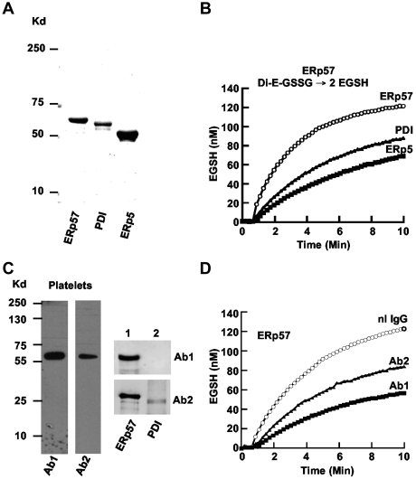
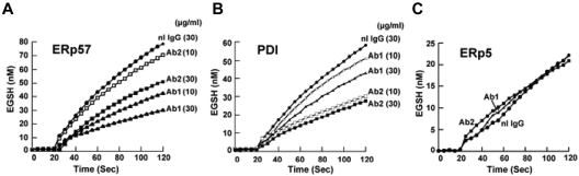
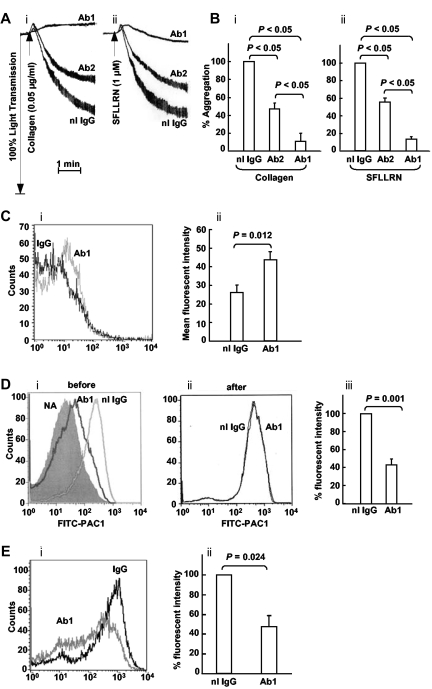
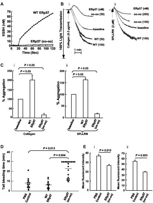
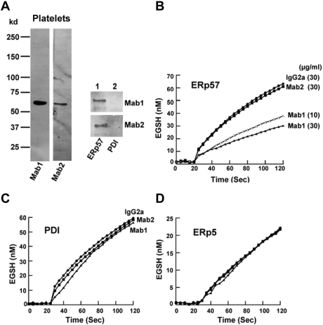
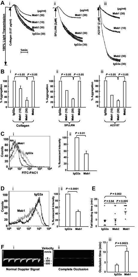
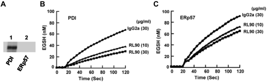
A close homologue to protein disulfide isomerase (PDI) called ERp57 forms disulfide bonds in glycoproteins in the endoplasmic reticulum and is expressed on the platelet surface. We generated 2 rabbit Abs to ERp57. One Ab strongly inhibited ERp57 in a functional assay and strongly inhibited platelet aggregation. There was minimal cross-reactivity of this Ab with PDI by Western blot or in the functional assay. This Ab substantially inhibited activation of the αIIbβ3 fibrinogen receptor and P-selectin expression. Furthermore, adding ERp57 to platelets potentiated aggregation. In contrast, adding a catalytically inactive ERp57 inhibited platelet aggregation. When infused into mice the inactive ERp57 prolonged the tail bleeding times. We generated 2 IgG2a mAbs that reacted with ERp57 by immunoblot. One of these Abs inhibited both ERp57 activity and platelet aggregation. The other Ab did not inhibit ERp57 activity or platelet aggregation. The inhibitory Ab inhibited activation of αIIbβ3 and P-selectin expression, prolonged tail bleeding times, and inhibited FeCl(3)-induced thrombosis in mice. Finally, we found that a commonly used mAb to PDI also inhibited ERp57 activity. We conclude that a glycoprotein-specific member of the PDI family, ERp57, is required for platelet aggregation, hemostasis, and thrombosis.


Figures

Figure 1
Figure 1
Comparison of purified ERp57, PDI, and ERp5 and characterization of rabbit anti-ERp57 Abs. (A) The relative sizes of the 3 enzymes on a gel under reducing conditions. The enzymes were expressed in E coli and purified by the His tag using a nickel column. (B) The enzymatic activities in the GSSG assay. A total of 150nM Di-E-GSSG was incubated in the presence of ERp57, PDI, or ERp5. The amount of EGSH formed over time is shown. (C) The reactivity of rabbit Ab 1 (Ab1) and Ab 2 (Ab2) on Western blot of platelet lysate (4 × 10⁸ platelets/lane) and purified ERp57 (lane 1) or PDI (lane 2; 200 ng protein/lane). (D) The inhibitory effect of Ab1 and Ab2 against ERp57 in the Di-E-GSSG assay compared with a normal rabbit IgG control (nL IgG); 30 μg/mL of each Ab was used in these studies. For the curves in panels B and D, the enzyme was added at ∼ 60 seconds.
Figure 2
Figure 2
Differential inhibition of purified ERp57 and PDI by rabbit anti-ERp57 Ab 1 and Ab 2. (A-C) The activity of ERp57, PDI, or ERp5 in the presence of Ab 1 (Ab1) and Ab 2 (Ab2) at 10 μg/mL or 30 μg/mL, or normal rabbit IgG (nL IgG) at 30 μg/mL. The amount of EGSH (nM) formed from Di-E-GSSG in the presence of enzyme over 120 seconds is shown. Each point is the composite of at least 3 samples (nL IgG, ●; Ab1 10 μg/mL, ▵; Ab1 30 μg/mL, ▴; Ab2 10 μg/mL, □; Ab2 30 μg/mL, ■). Inhibition was not increased with 60 μg/mL of either Ab (not shown). For these curves, the enzyme was added at ∼ 20 seconds.
Figure 3
Figure 3
The rabbit anti-ERP57 Ab 1 preferentially inhibits platelet aggregation. (A) The effect of Ab 1 and 2 are seen in representative tracings of platelet aggregation induced with (i) collagen and (ii) SFLLRN. (B) The combined data ± SE of at least 3 independent experiments is shown for collagen and SFLLRN. (C) The binding of Ab1 to resting platelets relative to normal IgG (i) with the cumulative results ± SE of a gated population of platelets also shown (ii; n = 8). (D) Anti-ERp57 (Ab1) inhibits SFLLRN (10μM)–induced PAC1 binding when added before (i), but not after (ii), activation. (i) PAC1 binding to nonactivated platelets (NA) is seen on the left. (iii) Inhibition of PAC1 binding by Ab1 as the percentage of fluorescent intensity relative to the normal rabbit IgG control, ± SE (n = 4). (E) A representative histogram (i) and combined data (ii) show that Ab1 inhibits P-selectin binding to convulxin (10 ng/mL)–activated platelets relative to normal IgG ± SE (n = 5). In these experiments, the Abs (30 μg/mL) were incubated with the platelets for 10 minutes before the addition of the agonist.
Figure 4
Figure 4
Wild-type ERp57 potentiates platelet aggregation and an inactive mutant ERp57 inhibits aggregation, prolongs the tail bleeding time in mice, and inhibits activation of αIIbβ3 and P-selectin expression ex vivo. (A) ERp57 with the 4 active site cysteines mutated to serine (oo-oo) is catalytically inactive. (B) Submaximal aggregation (baseline) was stimulated with collagen or the SFLLRN peptide. The potentiating effect of preincubating the platelets with purified wild-type ERp57 (WT ERp57) in the concentrations indicated is seen. The inhibitory (i) effect of adding the inactive (ii) mutant ERp57 (oo-oo) in the concentrations indicated is also seen. (C) The cumulative data ± SE of at least 3 different experiments, showing potentiation of aggregation by WT ERp57 and inhibition by the mutant ERp57 (oo-oo) relative to the baseline aggregation (i, collagen; ii, SFLLRN). The concentration of WT or mutant ERp57 that provided maximal potentiation or inhibition of responses varied a little between experiments. Fifty to 100nM WT or mutant ERp57 generally gave maximal potentiation and inhibition responses with collagen; 100 to 200nM of WT or mutant ERp57 provided maximal responses with the SFLLRN peptide. In these studies the enzymes were added for 10 minutes before the addition of the agonist. (D) The PBS control, 100 μg of WT ERp57, or 100 μg of the mutant ERp57 (oo-oo) were infused into mice, and the tail bleeding times were recorded up to 30 minutes. Horizontal bars represent mean bleeding times. (E) Results of ex vivo studies of platelets prepared from mice infused with PBS (control) or inactive ERp57 (oo-oo). The platelets were activated with convulxin (500 ng/mL) and activation of (i) αIIbβ3 (measured by binding of the JON/A Ab) and (ii) P-selectin expression were determined (n = 3).
Figure 5
Figure 5
Characterization of 2 monoclonal IgG2a Abs raised to ERp57. (A) The Western blots of the Abs called Mab1 and Mab2 against human platelet lysate (4 × 10⁸ platelets/lane) and purified ERp57 or PDI (200 μg protein/lane). Differential effect of the 2 IgG2a monoclonal anti-ERp57 Abs on (B) ERp57 activity and (C) lack of cross-reactivity to PDI or (D) ERp5 in the GSSG assay. For these studies, the activity of ERp57, PDI, and ERp5 alone (not shown in curves) was identical to enzyme with 30 μg/mL of the control IgG2a. A total of 30 μg/mL of each Ab was used in these studies except for ERp57 (B) where 10 μg/mL of the inhibitory Mab1 Ab was also tested. A total of 60 μg/mL of Mab 1 did not increase the inhibition (not shown). IgG2a control, ●; Mab1 10μg/mL, ▵; Mab1 30 μg/mL, ▴; Mab2 30 μg/mL, ■). (D) The curves for ERp5 with IgG2a, Mab1, or Mab2 (30 μg/mL of each Ab was used) completely overlapped. Each point is the composite of at least 3 samples using the Di-E-GSSG assay. (B-D) The enzyme was added at ∼ 20 seconds.
Figure 6
Figure 6
The inhibitory monoclonal anti-ERp57 Ab inhibits platelet aggregation, activation of αIIbβ3, P-selectin expression, and prolongs the tail bleeding times and the time to thrombosis in mice. (A) Representative tracings show Mab1 but not Mab2 inhibits aggregation of human platelets stimulated with collagen (i), the peptide SFLLRN (ii), or calcium ionophore (ii; A23187). (B) The combined data ± SE for at least 3 separate experiments (for collagen (i) and SFLLRN (ii), Mab1 at 10 μg/mL also inhibited aggregation to a P < .05 compared with either the IgG2a or Mab2 controls.) Calcium ionophore (iii) was used with platelets preincubated with apyrase (10 U/mL) and MeSAMP (100μM). (Ci) Mab1 inhibits PAC1 binding to SFLLRN (1μM)–activated platelets. PAC1 binding to nonactivated platelets (NA) is seen on the left. (ii) The results of inhibition of PAC1 binding as the percentage of fluorescent intensity relative to the normal IgG2a control ± SE (n = 3). (Di) A representative histogram and (ii) the combined data show the inhibitory monoclonal anti-ERp57 Ab Mab1 inhibits P-selectin binding to convulxin (10 ng/mL)–activated platelets relative to normal IgG2a ± SE (n = 7). (A-D) The platelets were preincubated with the inhibitory mAb (Mab1), noninhibitory Ab (Mab2), or control mouse IgG2a at 30 μg/mL for 10 minutes before the addition of the agonist. (E) A total of 200 μg of each Ab was infused into mice and the tail bleeding time recorded up to 15 minutes. Horizontal bars represent mean bleeding times. (Fi) The systolic and diastolic mouse carotid artery blood velocity monitored in mm of blood per second (mm/s) using the small animal Doppler probe with the Visual Sonics Vevo2100 flowmeter. (ii) Complete occlusion of blood flow after treatment with FeCl3. (iii) Mab1 inhibits FeCl3-induced occlusion relative to the normal mouse IgG2a control (n = 8). In these experiments, the carotid artery was treated with 5% FeCl3 for 2 minutes as described in “Studies using FaCl2-induced thrombosis of the carotid artery. A total of 450 μg of the control IgG2a or Mab1 were infused immediately before the filter paper soaked in 5% FeCl3 was applied to the carotid artery.”
Figure 7
Figure 7
The monoclonal anti-PDI Ab RL90 cross-reacts with ERp57 in the Di-E-GSSG assay. (A) Lack of cross- reactivity by Western blot to ERp57. (B) The inhibition of PDI. (C) The inhibition of ERp57 by RL90, at 10 μg/mL and 30 μg/mL relative to an isotype specific monoclonal mouse IgG2a Ab (30 μg/mL); 60 μg/mL did not provide more inhibition of PDI or ERp57 with the curves overlapping with the curves for 30 μg/mL. The amount of EGSH formed over 120 seconds from Di-E-GSSG is shown with the enzyme added at ∼ 20 seconds.

Similar articles

Cited by

References

    1. Goldberger RF, Epstein CJ, Anfinsen CB. Acceleration of reactivation of reduced bovine pancreatic ribonuclease by a microsomal system from rat liver. J Biol Chem. 1963;238:628–635. - PubMed
    1. Venetianer P, Straub FB. The enzymic reactivation of reduced ribonuclease. Biochim Biophys Acta. 1963;67:166–168. - PubMed
    1. Essex DW, Li M. Protein disulphide isomerase mediates platelet aggregation and secretion. Br J Haematol. 1999;104(3):448–454. - PubMed
    1. Essex DW. Redox control of platelet function. Antioxid Redox Signal. 2009;11(5):1191–1225. - PubMed
    1. Cho J, Furie BC, Coughlin SR, Furie B. A critical role for extracellular protein disulfide isomerase during thrombus formation in mice. J Clin Invest. 2008;118(3):1123–1131. - PMC - PubMed

Publication types

MeSH terms