. 2012 Feb 24;287(9):6679-92.
doi: 10.1074/jbc.M111.284232. Epub 2011 Dec 29.

The ubiquitin regulatory X (UBX) domain-containing protein TUG regulates the p97 ATPase and resides at the endoplasmic reticulum-golgi intermediate compartment

Affiliations

The ubiquitin regulatory X (UBX) domain-containing protein TUG regulates the p97 ATPase and resides at the endoplasmic reticulum-golgi intermediate compartment

Charisse M Orme et al. J Biol Chem. .

Abstract

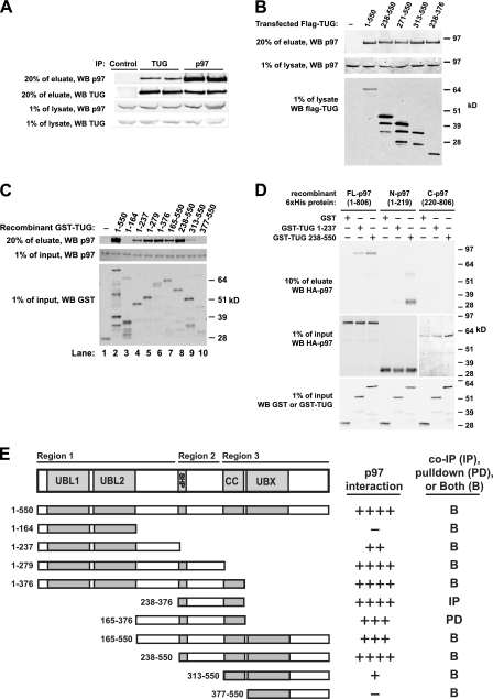
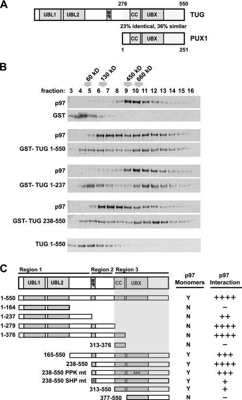
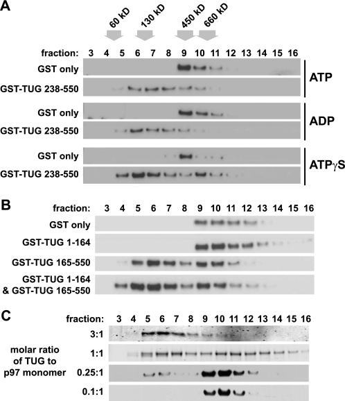
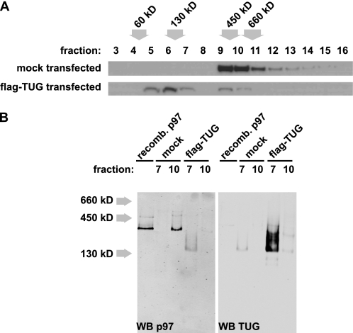
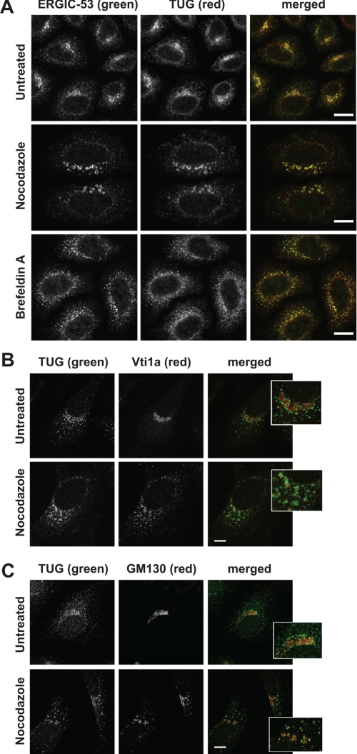
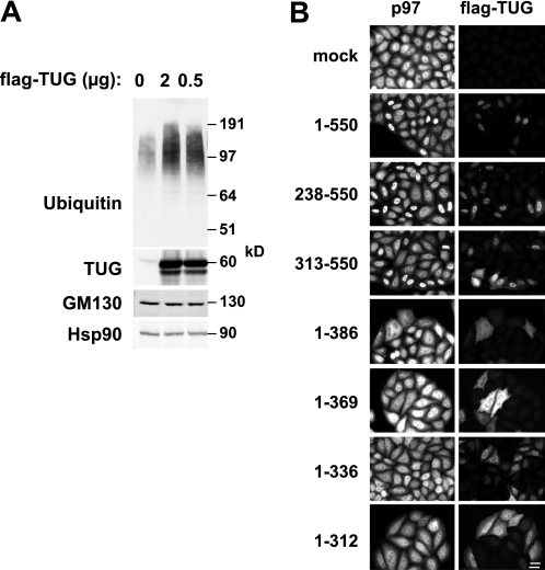
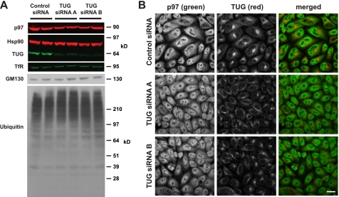
p97/VCP is a hexameric ATPase that is coupled to diverse cellular processes, such as membrane fusion and proteolysis. How p97 activity is regulated is not fully understood. Here we studied the potential role of TUG, a widely expressed protein containing a UBX domain, to control mammalian p97. In HEK293 cells, the vast majority of TUG was bound to p97. Surprisingly, the TUG UBX domain was neither necessary nor sufficient for this interaction. Rather, an extended sequence, comprising three regions of TUG, bound to the p97 N-terminal domain. The TUG C terminus resembled the Arabidopsis protein PUX1. Similar to the previously described action of PUX1 on AtCDC48, TUG caused the conversion of p97 hexamers into monomers. Hexamer disassembly was stoichiometric rather than catalytic and was not greatly affected by the p97 ATP-binding state or by TUG N-terminal regions in vitro. In HeLa cells, TUG localized to the endoplasmic reticulum-to-Golgi intermediate compartment and endoplasmic reticulum exit sites. Although siRNA-mediated TUG depletion had no marked effect on total ubiquitylated proteins or p97 localization, TUG overexpression caused an accumulation of ubiquitylated substrates and targeted both TUG and p97 to the nucleus. A physiologic role of TUG was revealed by siRNA-mediated depletion, which showed that TUG is required for efficient reassembly of the Golgi complex after brefeldin A removal. Together, these data support a model in which TUG controls p97 oligomeric status at a particular location in the early secretory pathway and in which this process regulates membrane trafficking in various cell types.


Figures

FIGURE 1.
FIGURE 1.
Interaction of TUG and p97. A, anti-TUG or anti-p97 antibodies were used to immunoprecipitate (IP) endogenous TUG and p97 from HEK293 cells lysed using 0.5% Nonidet P-40. Control IgG was used in lane 1, and duplicates of TUG (lanes 2 and 3) and p97 (lanes 4 and 5) immunoprecipitates are shown. Eluates and preimmunoprecipitation lysates were immunoblotted to detect TUG and p97, as indicated. B, HEK293 cells were transfected to express FLAG-tagged TUG proteins containing the indicated residues and lysed using 0.5% Nonidet P-40, and immunoprecipitations were performed using an anti-FLAG affinity matrix. Endogenous p97 was immunoblotted in eluates and pre-immunoprecipitation lysates, as indicated. C, recombinant GST or GST-TUG proteins containing the indicated TUG residues were used to pull down recombinant His6-p97 in 0.5% Nonidet P-40 buffer. Eluates and input proteins were immunoblotted using anti-p97 and anti-GST antibodies as indicated. D, GST or GST-TUG proteins were used to pull down His6-HA-tagged full-length, N-terminal, or C-terminal p97 proteins in 0.5% Nonidet P-40 buffer. Eluates and inputs were immunoblotted using anti-GST and anti-HA antibodies as indicated. Residues present in each p97 and TUG protein are indicated. E, diagram summarizes binding of p97 to TUG proteins containing the indicated residues. For reference, the overall TUG domain structure is shown at top. The relative amount of p97 that associated with each TUG protein is indicated at the right. Binding was tested using both GST pull-downs (PD; as in C and D) and by immunoprecipitation (IP; as in B). Most interactions were tested both ways, as indicated (B), and similar results were obtained using both methods. Each construct was tested at least twice, with most tested three times. WB, Western blot.
FIGURE 2.
FIGURE 2.
TUG generates p97 monomers. A, diagrams of the domain structures of TUG and PUX1; a region of similarity is indicated. The full alignment is shown in supplemental Fig. 1. B, recombinant His6-p97 proteins were incubated with GST alone or with the indicated GST-TUG proteins in equimolar amounts. After 30 min at 4 °C, p97 complexes were separated by sedimentation on sucrose gradients and analyzed by immunoblotting for p97 and TUG, as indicated. Fractions are numbered from the top of the gradient, and the positions of molecular weight standards are indicated above the fraction numbers. The position on the gradient of TUG alone is shown at the bottom. C, the diagrammed TUG proteins were expressed as recombinant GST fusions and were tested for their ability to generate p97 monomers using sedimentation. A 3:1 molar ratio of TUG/p97 proteins was used. All constructs were tested at least twice. The shaded area corresponds to the region of TUG that was necessary for generation of p97 monomers. For reference, the relative ability of each TUG construct to copurify p97 in pull-down and/or coimmunoprecipitation experiments is shown at the right.
FIGURE 3.
FIGURE 3.
p97 hexamer disassembly in vitro is not affected by nucleotides or by TUG N-terminal domains and requires stoichiometric amounts of TUG. A, recombinant His6-p97 was preincubated with ATP, ADP, or ATPγS at final concentrations of 1 mm for 10 min at 4 °C, and then GST or GST-TUG 238–550 was added, and the incubation was continued for an additional 30 min at 4 °C. The molar ratio of TUG to p97 was 3:1. p97 oligomeric status was analyzed by sedimentation on sucrose density gradients, followed by immunoblotting of the fractions as indicated. The positions of molecular weight markers are shown at the top. The experiment was repeated three times with similar results. B, recombinant His6-p97 was incubated for 30 min at 4 °C with GST alone or with GST TUG 1–164 and/or GST TUG 165–550, as indicated. Molar ratios of 3:1 (TUG/p97) were used. p97 complexes were then separated by sedimentation on sucrose gradients, and the fractions were immunoblotted to detect p97. The experiment was performed twice with similar results. C, GST-TUG 238–550 was incubated with His6-p97 at the indicated molar ratios of TUG/p97 monomer, and then complexes were sedimented on sucrose gradients. Fractions were immunoblotted to detect p97.
FIGURE 4.
FIGURE 4.
Transfected TUG generates and binds p97 monomers. A, HEK293 cells were transfected with FLAG-tagged TUG and lysed using 0.5% Nonidet P-40. Postnuclear supernatants were analyzed by sedimentation on sucrose density gradients, and fractions were immunoblotted to detect p97. The positions of molecular weight standards are indicated. The experiment was repeated with similar results. B, proteins in fractions 7 and 10 from the gradients in A were separated using native PAGE and then immunoblotted as indicated. Recombinant His6-p97 was included in the first lane for comparison, and the position of molecular weight standards is indicated at the left. Western blots (WB) of p97 and TUG were done on the same membrane and were imaged at different wavelengths using a LI-COR Odyssey imaging system. The experiment was repeated with similar results.
FIGURE 5.
FIGURE 5.
TUG colocalizes with ERGIC-53 by confocal microscopy. A, endogenous TUG and ERGIC-53 were detected in HeLa cells using indirect immunofluorescence and confocal microscopy. Cells were untreated or treated with 30 μm nocodazole or 10 μg/ml brefeldin A for 2 h prior to staining, as indicated. B and C, endogenous TUG and Vti1a (B) and TUG and GM130 (C) were imaged as in A. The insets show enlargements of the perinuclear region of the merged images. Scale bars, 10 μm.
FIGURE 6.
FIGURE 6.
TUG overexpression affects ubiquitylated protein abundance and p97 distribution. A, HeLa cells were transfected using the indicated amounts of a plasmid to express full-length, FLAG-tagged TUG. Cells were lysed 48 h after transfection, and immunoblots were done to detect the indicated proteins. An antibody to the TUG C terminus was used to detect endogenous and transfected TUG and to assess the level of overexpression. B, FLAG-tagged intact TUG (residues 1–550) or truncated TUG proteins (containing the indicated residues) were transfected HeLa cells. Cells were fixed, and immunofluorescence microscopy was performed to detect endogenous p97 and FLAG-tagged TUG, as indicated. Scale bar, 20 μm.
FIGURE 7.
FIGURE 7.
siRNA-mediated TUG depletion has no large effect on ubiquitylated protein abundance or p97 distribution. A, HeLa were transfected using control (scrambled) siRNA, TUG siRNA A, or TUG siRNA B, as indicated, and were lysed 48 h after transfection. Proteins were separated by SDS-PAGE and immunoblotted as indicated. Hsp90 and transferrin receptor were used as loading controls. B, endogenous TUG and p97 were imaged in HeLa cells using indirect immunofluorescence and confocal microscopy. Cells were treated with the indicated siRNAs 48 h prior to fixation. Scale bar, 20 μm.
FIGURE 8.
FIGURE 8.
TUG depletion alters GM130 staining intensity and impairs Golgi reformation after brefeldin A washout. A, endogenous TUG and GM130 were imaged using immunofluorescence and confocal microscopy of HeLa cells. Cells were treated with control siRNA, TUG siRNA A, or TUG siRNA B, as indicated, for 48 h prior to fixation. B, HeLa cells were treated with control siRNA or TUG siRNA A for 48 h and then mock-treated or treated with BFA, as indicated. BFA was used at 10 μg/ml at 37 °C for 1 h, and then cells were washed three times and allowed to recover in the absence of BFA for 1 h at 37 °C. Cells were then fixed and stained to detect GM130 as in A. The experiment was repeated with similar results. C, images of cells in B were quantified. For each cell, the ratio of fluorescence intensity in the perinuclear region to that of the entire cell was quantified. 19–24 cells were analyzed per condition, and the mean ratio ± S.E. (error bars) is plotted. Statistical significance was assessed using a two-tailed t test. Scale bars, 20 μm.

References

    1. White S. R., Lauring B. (2007) AAA+ ATPases. Achieving diversity of function with conserved machinery. Traffic 8, 1657–1667 - PubMed
    1. Brunger A. T., DeLaBarre B. (2003) NSF and p97/VCP. Similar at first, different at last. FEBS Lett. 555, 126–133 - PubMed
    1. Schuberth C., Buchberger A. (2008) UBX domain proteins. Major regulators of the AAA ATPase Cdc48/p97. Cell Mol. Life Sci. 65, 2360–2371 - PMC - PubMed
    1. Vij N. (2008) AAA ATPase p97/VCP. Cellular functions, disease and therapeutic potential. J. Cell Mol. Med. 12, 2511–2518 - PMC - PubMed
    1. Yeung H. O., Kloppsteck P., Niwa H., Isaacson R. L., Matthews S., Zhang X., Freemont P. S. (2008) Insights into adaptor binding to the AAA protein p97. Biochem. Soc. Trans. 36, 62–67 - PubMed

Publication types

MeSH terms