

Optimization of a 96-Well Electroporation Assay for Postnatal Rat CNS Neurons Suitable for Cost-Effective Medium-Throughput Screening of Genes that Promote Neurite Outgrowth
- PMID: 22207835
- PMCID: PMC3245668
- DOI: 10.3389/fnmol.2011.00055
Optimization of a 96-Well Electroporation Assay for Postnatal Rat CNS Neurons Suitable for Cost-Effective Medium-Throughput Screening of Genes that Promote Neurite Outgrowth
Abstract
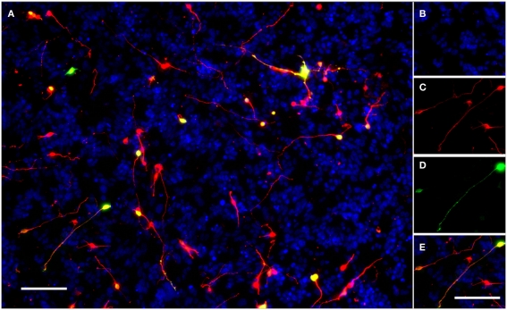
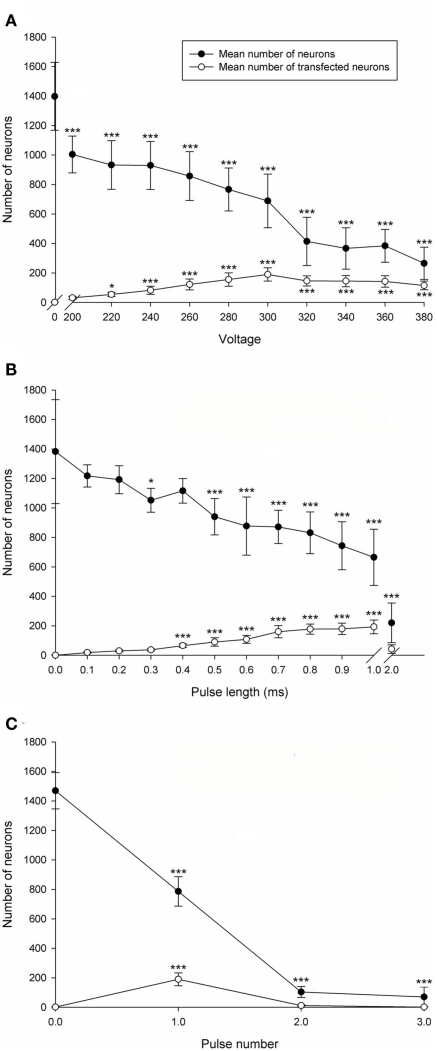
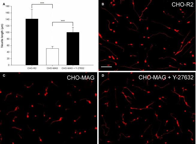
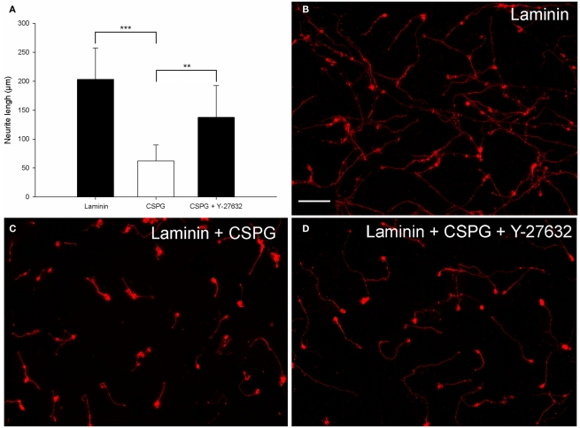
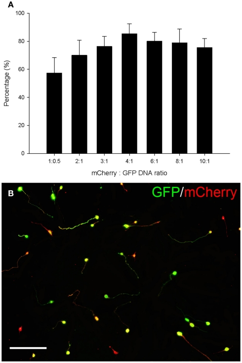
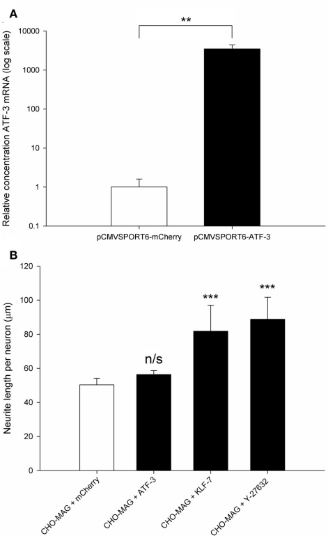
Following an injury, central nervous system (CNS) neurons show a very limited regenerative response which results in their failure to successfully form functional connections with their original target. This is due in part to the reduced intrinsic growth state of CNS neurons, which is characterized by their failure to express key regeneration-associated genes (RAGs) and by the presence of growth inhibitory molecules in CNS environment that form a molecular and physical barrier to regeneration. Here we have optimized a 96-well electroporation and neurite outgrowth assay for postnatal rat cerebellar granule neurons (CGNs) cultured upon an inhibitory cellular substrate expressing myelin-associated glycoprotein or a mixture of growth inhibitory chondroitin sulfate proteoglycans. Optimal electroporation parameters resulted in 28% transfection efficiency and 51% viability for postnatal rat CGNs. The neurite outgrowth of transduced neurons was quantitatively measured using a semi-automated image capture and analysis system. The neurite outgrowth was significantly reduced by the inhibitory substrates which we demonstrated could be partially reversed using a Rho Kinase inhibitor. We are now using this assay to screen large sets of RAGs for their ability to increase neurite outgrowth on a variety of growth inhibitory and permissive substrates.
Keywords: 96-well; CNS neuron; RAG; electroporation; inhibitory substrate; neurite outgrowth; neuronal regeneration; transfection.
Figures
Similar articles
-
A quantitative method for analysis of in vitro neurite outgrowth.J Neurosci Methods. 2007 Aug 30;164(2):350-62. doi: 10.1016/j.jneumeth.2007.04.021. Epub 2007 May 6. J Neurosci Methods. 2007. PMID: 17570533
-
Neuronal age influences the response to neurite outgrowth inhibitory activity in the central and peripheral nervous systems.Brain Res. 1999 Jul 31;836(1-2):49-61. doi: 10.1016/s0006-8993(99)01588-7. Brain Res. 1999. PMID: 10415404
-
Epac2 Elevation Reverses Inhibition by Chondroitin Sulfate Proteoglycans In Vitro and Transforms Postlesion Inhibitory Environment to Promote Axonal Outgrowth in an Ex Vivo Model of Spinal Cord Injury.J Neurosci. 2019 Oct 16;39(42):8330-8346. doi: 10.1523/JNEUROSCI.0374-19.2019. Epub 2019 Aug 13. J Neurosci. 2019. PMID: 31409666 Free PMC article.
-
Neurite outgrowth inhibitors in gliotic tissue.Adv Exp Med Biol. 1999;468:207-24. doi: 10.1007/978-1-4615-4685-6_17. Adv Exp Med Biol. 1999. PMID: 10635031 Review.
-
Insights into the physiological role of CNS regeneration inhibitors.Front Mol Neurosci. 2015 Jun 11;8:23. doi: 10.3389/fnmol.2015.00023. eCollection 2015. Front Mol Neurosci. 2015. PMID: 26113809 Free PMC article. Review.
Cited by
-
Overexpression of the Fibroblast Growth Factor Receptor 1 (FGFR1) in a Model of Spinal Cord Injury in Rats.PLoS One. 2016 Mar 25;11(3):e0150541. doi: 10.1371/journal.pone.0150541. eCollection 2016. PLoS One. 2016. PMID: 27015635 Free PMC article.
-
The use of an adeno-associated viral vector for efficient bicistronic expression of two genes in the central nervous system.Methods Mol Biol. 2014;1162:189-207. doi: 10.1007/978-1-4939-0777-9_16. Methods Mol Biol. 2014. PMID: 24838969 Free PMC article.
-
Physical non-viral gene delivery methods for tissue engineering.Ann Biomed Eng. 2013 Mar;41(3):446-68. doi: 10.1007/s10439-012-0678-1. Epub 2012 Oct 26. Ann Biomed Eng. 2013. PMID: 23099792 Free PMC article. Review.
-
Axonal Regeneration after Spinal Cord Injury: Molecular Mechanisms, Regulatory Pathways, and Novel Strategies.Biology (Basel). 2024 Sep 7;13(9):703. doi: 10.3390/biology13090703. Biology (Basel). 2024. PMID: 39336130 Free PMC article. Review.
-
The Adaptor Protein CD2AP Is a Coordinator of Neurotrophin Signaling-Mediated Axon Arbor Plasticity.J Neurosci. 2016 Apr 13;36(15):4259-75. doi: 10.1523/JNEUROSCI.2423-15.2016. J Neurosci. 2016. PMID: 27076424 Free PMC article.
References
LinkOut - more resources
Full Text Sources