

Time determines the neural circuit underlying associative fear learning
- PMID: 22207842
- PMCID: PMC3246300
- DOI: 10.3389/fnbeh.2011.00089
Time determines the neural circuit underlying associative fear learning
Abstract
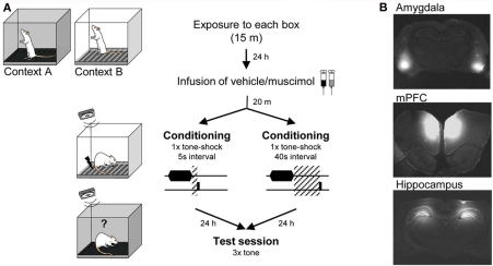
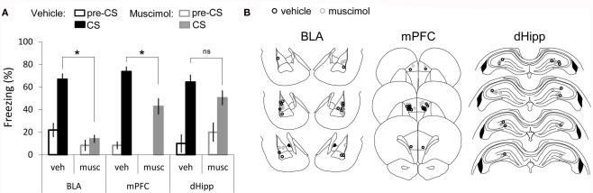
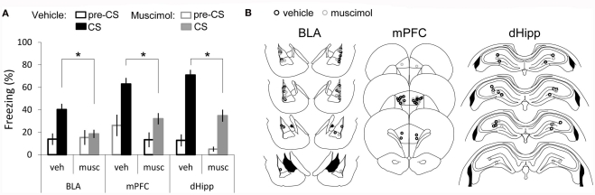
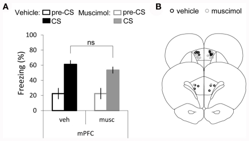
Ultimately associative learning is a function of the temporal features and relationships between experienced stimuli. Nevertheless how time affects the neural circuit underlying this form of learning remains largely unknown. To address this issue, we used single-trial auditory trace fear conditioning and varied the length of the interval between tone and foot-shock. Through temporary inactivation of the amygdala, medial prefrontal-cortex (mPFC), and dorsal-hippocampus in rats, we tested the hypothesis that different temporal intervals between the tone and the shock influence the neuronal structures necessary for learning. With this study we provide the first experimental evidence showing that temporarily inactivating the amygdala before training impairs auditory fear learning when there is a temporal gap between the tone and the shock. Moreover, imposing a short interval (5 s) between the two stimuli also relies on the mPFC, while learning the association across a longer interval (40 s) becomes additionally dependent on a third structure, the dorsal-hippocampus. Thus, our results suggest that increasing the interval length between tone and shock leads to the involvement of an increasing number of brain areas in order for the association between the two stimuli to be acquired normally. These findings demonstrate that the temporal relationship between events is a key factor in determining the neuronal mechanisms underlying associative fear learning.
Keywords: amygdala; hippocampus; mPFC; muscimol; single-trial; trace fear conditioning.
Figures
Similar articles
-
Time-dependent involvement of the dorsal hippocampus in trace fear conditioning in mice.Hippocampus. 2005;15(4):418-26. doi: 10.1002/hipo.20067. Hippocampus. 2005. PMID: 15669102
-
A prefrontal-bed nucleus of the stria terminalis circuit limits fear to uncertain threat.Elife. 2020 Dec 15;9:e60812. doi: 10.7554/eLife.60812. Elife. 2020. PMID: 33319747 Free PMC article.
-
Re-emergence of extinguished auditory-cued conditioned fear following a sub-conditioning procedure: effects of hippocampal and prefrontal tetanic stimulations.Neurobiol Learn Mem. 2011 May;95(4):510-8. doi: 10.1016/j.nlm.2011.03.002. Epub 2011 Mar 21. Neurobiol Learn Mem. 2011. PMID: 21397708
-
Distributed coding in auditory thalamus and basolateral amygdala upon associative fear learning.Curr Opin Neurobiol. 2021 Apr;67:183-189. doi: 10.1016/j.conb.2020.11.014. Epub 2020 Dec 26. Curr Opin Neurobiol. 2021. PMID: 33373858 Review.
-
Role of the basolateral amygdala and NMDA receptors in higher-order conditioned fear.Rev Neurosci. 2011;22(3):317-33. doi: 10.1515/RNS.2011.025. Epub 2011 May 13. Rev Neurosci. 2011. PMID: 21591909 Review.
Cited by
-
NR2A- and NR2B-containing NMDA receptors in the prelimbic medial prefrontal cortex differentially mediate trace, delay, and contextual fear conditioning.Learn Mem. 2013 May 15;20(6):290-4. doi: 10.1101/lm.030510.113. Learn Mem. 2013. PMID: 23676200 Free PMC article.
-
Functional network of contextual and temporal memory has increased amygdala centrality and connectivity with the retrosplenial cortex, thalamus, and hippocampus.Sci Rep. 2023 Aug 11;13(1):13087. doi: 10.1038/s41598-023-39946-1. Sci Rep. 2023. PMID: 37567967 Free PMC article.
-
Deletion of Dtnbp1 in mice impairs threat memory consolidation and is associated with enhanced inhibitory drive in the amygdala.Transl Psychiatry. 2019 Apr 9;9(1):132. doi: 10.1038/s41398-019-0465-y. Transl Psychiatry. 2019. PMID: 30967545 Free PMC article.
-
Sound check, stage design and screen plot - how to increase the comparability of fear conditioning and fear extinction experiments.Psychopharmacology (Berl). 2019 Jan;236(1):33-48. doi: 10.1007/s00213-018-5111-5. Epub 2018 Nov 23. Psychopharmacology (Berl). 2019. PMID: 30470861 Free PMC article. Review.
-
Apoaequorin differentially modulates fear memory in adult and aged rats.Brain Behav. 2020 Nov;10(11):e01832. doi: 10.1002/brb3.1832. Epub 2020 Sep 18. Brain Behav. 2020. PMID: 32945630 Free PMC article.
References
LinkOut - more resources
Full Text Sources
Other Literature Sources
Miscellaneous