. 2011 Jul;2(7):745-52.
doi: 10.1177/1947601911426007.

Caspase-2-Based Regulation of the Androgen Receptor and Cell Cycle in the Prostate Cancer Cell Line LNCaP

Affiliations

Caspase-2-Based Regulation of the Androgen Receptor and Cell Cycle in the Prostate Cancer Cell Line LNCaP

Agshin F Taghiyev et al. Genes Cancer. 2011 Jul.

Abstract

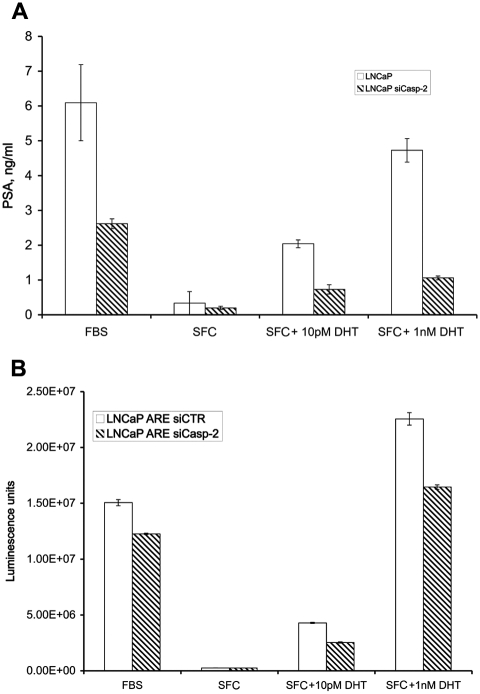
Caspase-2 can induce apoptosis in response to extrinsic and intrinsic signals. Unlike other caspases, this protein is not expressed solely in nonnuclear compartments; a subpopulation is constitutively localized in the nucleus. As one of the most evolutionarily conserved caspases, caspase-2 may have roles in multiple cellular processes. However, its contribution to nonapoptotic processes remains a mystery. In this study, we show that caspase-2 activity is important for proliferation by cells of the androgen-dependent prostate cancer cell line LNCaP. LNCaP cells expressing either a dominant-negative (dn) form of caspase or an siRNA against caspase-2 had lower androgen receptor (AR)-dependent proliferative responses than control cells, and application of the siRNA resulted in downregulation of the expression of both AR-dependent prostate-specific antigen (PSA) and AR-dependent reporter luciferase. Also, caspase-2 formed complexes with the cell cycle regulatory proteins cyclin D3, CDK4, and p21/Cip1, and caspase-2 regulated AR transactivation by inhibiting the repressive function of cyclin D3. Taken together, these results reveal, for the first time, that caspase-2 is involved in cell cycle promotion and AR activation. Given that prostate cancer cells depend on AR activity in order to survive, the fact that our data indicate that caspase-2 positively regulates AR activity suggests that caspase-2 has potential as a target in the treatment of prostate cancer.

Keywords: CDK4; DHT; LNCaP; apoptosis; caspase-2; cell cycle; cyclin D3; p21/Cip1; prostate cancer.


Conflict of interest statement

The author(s) declared no potential conflicts of interest with respect to the research, authorship, and/or publication of this article.

Figures

Figure 1.
Figure 1.
The proliferative response to DHT in LNCaP is caspase-2 dependent. (A) Thymidine uptake in LNCaP-Puro, LNCaP Caspase-2dn, and LNCaP siCaspase-2 cells that were plated at a density of 7,000 cells/well in 96-well flat-bottomed plates and cultured under SFC alone for 72 hours. DHT was added for 48 hours at the indicated concentrations. Each bar represents the mean value of three replicates, in one of four independent experiments, and bars represent standard error. The proliferative response was estimated as described in Materials and Methods, based on [3H] thymidine uptake. (B) Cell number in LNCaP-Puro, LNCaP Caspase-2dn, and LNCaP siCaspase-2 cells plated at a density of 30,000 cells/well in a 12-well plate and cultured under SFC alone for 96 hours. There was 10 pM DHT added for 72 hours. Each bar represents mean values of three counts from two replicates in one of two independent experiments, and bars represent standard error.
Figure 2.
Figure 2.
Caspase-2 regulates AR transactivation. (A) PSA levels in the culture medium. LNCaP-Hygro and LNCaP siCaspase-2 cells were cultured in 24-well plates for 48 hours under SFC and then treated for 24 hours with indicated concentrations of DHT. PSA levels in the culture medium were estimated using the Free PSA ELISA kit, as described in Materials and Methods. The amount of PSA was calculated using a calibration curve based on varying PSA concentrations and was normalized to the number of cells per well. Each bar represents the mean value of two replicates for one of three independent experiments, and bars represent standard error. (B) Luminescence intensity in cell lysates. LNCaP-ARE-siCTR and LNCaP-ARE-siCaspase-2 cells were cultured in 24-well plates for 48 hours under SFC and then treated for 24 hours with DHT at the indicated concentrations. Luminescence intensity in cell lysates was estimated using a luminometer as described in Materials and Methods. Each bar represents the mean value of two measurements in one of three independent experiments, and bars represent standard error.
Figure 3.
Figure 3.
Caspase-2 regulates the AR–cyclin D3 interaction. Equal amounts of cell lysates from untreated LNCaP-siCTR and LNCaP siCaspase-2 cells were subjected to immunoprecipitation analysis using mouse anti-AR and mouse anti–cyclin D3 antibodies. The same antibodies were used to visualize proteins by Western blot analysis. The figure is representative of three experiments.
Figure 4.
Figure 4.
Caspase-2 forms a complex with cyclin D3, CDK4, and p21/Cip1. (A) Immunoprecipitation analysis. LNCaP cells were cultured under SFC alone for 72 hours and then treated with 10 pM DHT for 72 hours. Cell lysates were subjected to immunoprecipitation analysis using rabbit anti–caspase-2 antibodies as described in Materials and Methods. Western blotting was carried out using antibody indicated to the right (see Materials and Methods). Blot is representative of three experiments. (B) Assessments of caspase-2 and p21/Cip1 levels. LNCaP-Hygro and LNCaP siCaspase-2 cells incubated in SFC alone for 72 hours and then treated with 10 pM DHT for 72 and 120 hours. Total cell lysates of treated and untreated cells were prepared as described in Materials and Methods. Caspase-2 and p21/Cip1 protein levels were assessed by Western blot analysis. β-actin was used as loading control. Blot shown is representative of three separate experiments.
Figure 5.
Figure 5.
TSA-induced formation of caspase-2/cyclin D3/p21 complexes inhibits HDAC activity. Immunoprecipitation analysis. (A) LNCaP and (B) Du145 cells were treated with 100 ng/mL TSA for 16 to 20 hours. Immunoprecipitation analysis of cell lysates was carried out using rabbit anti–caspase-2 antibodies as described in Materials and Methods. Proteins levels were assessed by Western blotting with antibodies indicated to the right of the blot (see Materials and Methods). Panels each represent one of four experiments with similar results.
Figure 6.
Figure 6.
Links between caspase-2 and cell signaling pathways that regulate apoptosis and the cell cycle. Critical elements of the suggested model are schematically represented (see details in the Discussion).

Similar articles

Cited by

References

    1. Zhivotovsky B, Orrenius S. Caspase-2 function in response to DNA damage. Biochem Biophys Res Commun. 2005;331:859-67 - PubMed
    1. Fuentes-Prior P, Salvesen GS. The protein structures that shape caspase activity, specificity, activation and inhibition. Biochem J. 2004;384:201-32 - PMC - PubMed
    1. Tinel A, Tschopp J. The PIDDosome, a protein complex implicated in activation of caspase-2 in response to genotoxic stress. Science. 2004;304:843-6 - PubMed
    1. Baliga BC, Read SH, Kumar S. The biochemical mechanism of caspase-2 activation. Cell Death Differ. 2004;11:1234-41 - PubMed
    1. Mancini M, Machamer CE, Roy S, et al. Caspase-2 is localized at the Golgi complex and cleaves golgin-160 during apoptosis. J Cell Biol. 2000;149:603-12 - PMC - PubMed

LinkOut - more resources