. 2011 Oct;42(4):259-65.
doi: 10.1177/155005941104200412.

A micro-electrocorticography platform and deployment strategies for chronic BCI applications

Affiliations

A micro-electrocorticography platform and deployment strategies for chronic BCI applications

Sanitta Thongpang et al. Clin EEG Neurosci. 2011 Oct.

Abstract

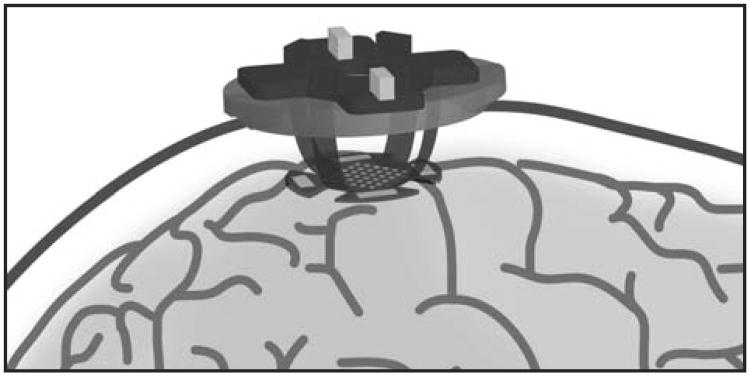
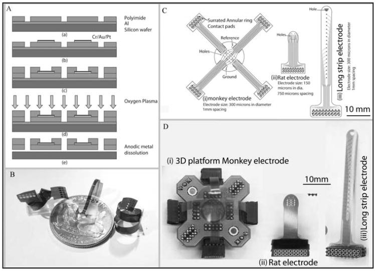
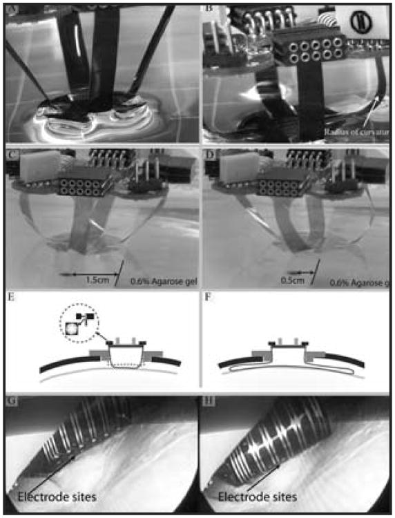
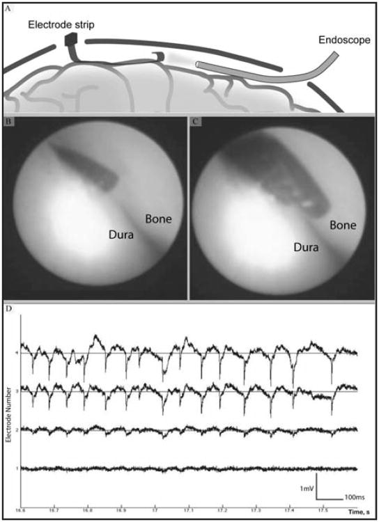
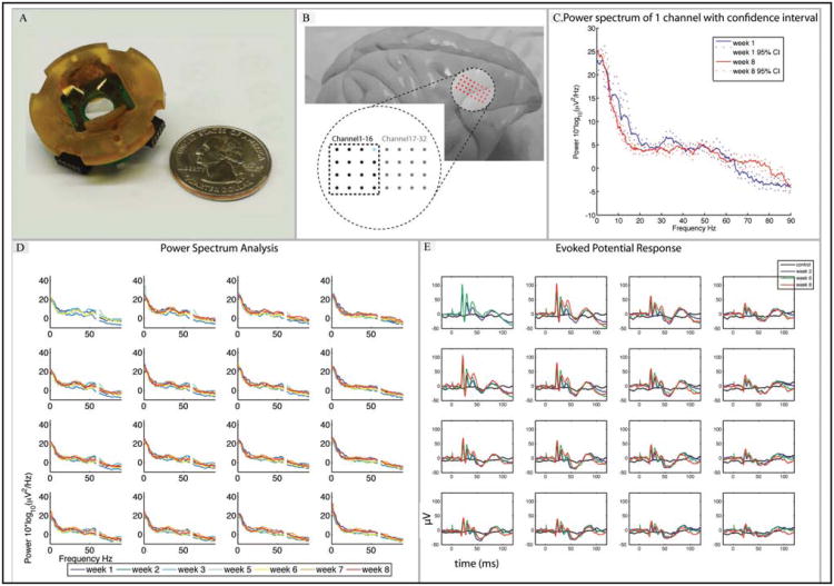
Over the past decade, electrocorticography (ECoG) has been used for a wide set of clinical and experimental applications. Recently, there have been efforts in the clinic to adapt traditional ECoG arrays to include smaller recording contacts and spacing. These devices, which may be collectively called "micro-ECoG" arrays, are loosely defined as intercranial devices that record brain electrical activity on the sub-millimeter scale. An extensible 3D-platform of thin film flexible micro-scale ECoG arrays appropriate for Brain-Computer Interface (BCI) application, as well as monitoring epileptic activity, is presented. The designs utilize flexible film electrodes to keep the array in place without applying significant pressure to the brain and to enable radial subcranial deployment of multiple electrodes from a single craniotomy. Deployment techniques were tested in non-human primates, and stimulus-evoked activity and spontaneous epileptic activity were recorded. Further tests in BCI and epilepsy applications will make the electrode platform ready for initial human testing.


Conflict of interest statement

Disclosure and Conflict of Interest: S. Thongpang, T.J. Richner, S.K. Brodnick, A. Schendel, J. Kim, J.A. Wilson, J. Hippensteel, L. Krugner-Higby, D. Moran, A.S. Ahmed, D. Neimann, K. Sillay, J.C. Williams have no conflicts of interest in relation to this article.

Figures

Figure 1
Figure 1
A schematic representation of the 3D-electrode platform on a primate brain with the alternative ground and reference pads shown surrounding the perimeter of the electrode array.
Figure 2
Figure 2
A) Electrode fabrication process using lithography to pattern Polyimide, metal evaporation to deposit the traces and electrode sites, and anodic metal dissolution to release the electrodes. B) Various electrode designs that show the flexibility of the polyimide polymer substrate, making it suitable for a wide range of applications. C) The layout of a variety of electrode designs with different shapes, electrode sizes and inter-electrode spacing. D) The realization of the design layout in (C) with corresponding platform and connectors.
Figure 3
Figure 3
A) Four legs allow the 2-D array to translate along 3 axes and rotate about an additional 2 axes. The flexible legs maintain only a minimal amount of pressure to ensure close contact between the electrode and tissue. B) The hydrophilicity of the polyimide helps the electrode adhere to the dura or pia mater. C) and D) An x- axis translation of the brain tissue phantom (0.6% agarose gel) up to 1 cm, representing the constant contact of the electrode array with dura due to small brain movement in all x-, y-, z- and 3 rotational axes in vivo. E) A cross-section of the electrode platform shows an assembled electrode and PCB sitting atop a plastic adapter ring inside a craniotomy. The adapter ring can be temporarily fixed to the cranium. The inset shows the multi-leafed electrode contact to the electrical sockets on the PCB. F) Diagram illustrating the flexible legs (longer leg cable than those shown in A-E), which flare out radially under the skull to cover a larger area of cortex. G) and H) Electrode sites come in contact with the dura as the electrode is deployed. The dura can be left intact (pictured) or removed.
Figure 4
Figure 4
Long strip electrode suitable for acute recording. A) A representation of a strip electrode inserted through a very small (10 mm) burr hole simultaneously with an endoscope for real time imaging of electrode position. B) and C) Real time images of electrodes in contact with the dura, taken from the scope which was situated on the opposite side of the location of electrode entry. D) Epileptic activity observed from an acute recording using the same long strip electrode array. Spontaneous epileptic activity recorded under anethesia from 4 neighboring electrode sites shows the signal amplitude of up to 2 mV, droping over a spatial scale resolvable only by micro-ECoG arrays.
Figure 5
Figure 5
Long-term micro-ECoG recording comparison. A) An electrode array and platform design that fits a 19 mm craniotomy in comparison with a quarter coin. B) Position of the electrode array on a non-human primate brain (Rhesus macaque), situated over sensorimotor cortex. C) Power spectrum (with 95% confidence intervals) of an example electrode site (highlighted in blue in (Figure 5B)) showing stability of the recorded baseline signal over an 8 week period. D) Power spectra of channels 1-16 over 8 weeks show long-term stability in long-term implant (60 Hz line noise omitted). E) Evoked potential response due to contralateral hind limb electrical stimulation (2.5 mA, 2 ms) compared to control (no stimulus) (week 2, 6 and 8).

References

    1. Hollenberg B, Richards C, Richards R, Bahr D, Rector D. A MEMS fabricated flexible electrode array for recording surface field potentials. J Neurosci Methods. 2006;153(1):147–153. - PubMed
    1. Rubehn B, Bosman C, Oostenveld R, Fries P, Stieglitz T. A MEMS-based flexible multichannel ECoG-electrode array. J Neural Eng. 2009;6(3):036003. - PubMed
    1. Viventi J, Kim D, Moss J, Kim Y, Blanco J, Annetta N, et al. A conformal, bio-interfaced class of silicon electronics for mapping cardiac electrophysiology. Sci Transl Med. 2010;2(24):24ra22. - PMC - PubMed
    1. Kellis S, House P, Thomson K, Brown R, Greger B. Human neocortical electrical activity recorded on nonpenetrating microwire arrays: applicability for neuroprostheses. Neurosurg Focus. 2009;27(1):E9. - PMC - PubMed
    1. Blanco J, Stead M, Krieger A, Viventi J, Marsh W, Lee K, et al. Unsupervised classification of high-frequency oscillations in human neocortical epilepsy and control patients. J Neurophysiol. 2010;104(5):2900–2912. - PMC - PubMed

Publication types