. 2011 Dec 21;101(12):2929-38.
doi: 10.1016/j.bpj.2011.11.009. Epub 2011 Dec 20.

Aqueous viscosity is the primary source of friction in lipidic pore dynamics

Affiliations

Aqueous viscosity is the primary source of friction in lipidic pore dynamics

Rolf Ryham et al. Biophys J. .

Abstract

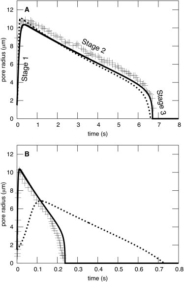
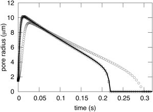
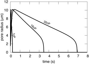
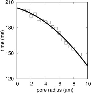
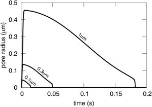
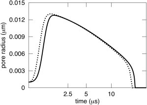
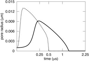
A new theory, to our knowledge, is developed that describes the dynamics of a lipidic pore in a liposome. The equations of the theory capture the experimentally observed three-stage functional form of pore radius over time--stage 1, rapid pore enlargement; stage 2, slow pore shrinkage; and stage 3, rapid pore closure. They also show that lipid flow is kinetically limited by the values of both membrane and aqueous viscosity; therefore, pore evolution is affected by both viscosities. The theory predicts that for a giant liposome, tens of microns in radius, water viscosity dominates over the effects of membrane viscosity. The edge tension of a lipidic pore is calculated by using the theory to quantitatively account for pore kinetics in stage 3, rapid pore closing. This value of edge tension agrees with the value as standardly calculated from the stage of slow pore closure, stage 2. For small, submicron liposomes, membrane viscosity affects pore kinetics, but only if the viscosity of the aqueous solution is comparable to that of distilled water. A first-principle fluid-mechanics calculation of the friction due to aqueous viscosity is in excellent agreement with the friction obtained by applying the new theory to data of previously published experimental results.


Figures

Figure 1
Figure 1
Pore evolution in giant liposomes exhibits three distinct stages. (A) Both BGS and DAV theory yield three stages, and parameters can be found for each theory to satisfactorily fit experimental data. But the curve for BGS (dashed curve) assumes ηs = 0 and ηl = 1000 P. The DAV model (solid curve), on the other hand, assumes ηs = 32 cP as used in the experiments of Brochard-Wyart et al. (10) (crosses), and the physically realistic ηl = 1 P. The other parameters were also as in Brochard-Wyart et al. (10) and are S = 0.0458 kT/nm2, γ = 2.5 kT/nm (∼10 pN), W = 0 kT/nm2, R0= 19.7 μm, R(t = 0) = 20.59 μm, r(t = 0) = 1.5 μm, C = 8.16, and d = 3 nm. (B) Parameters used are the same as in A, except that ηs = 1.13 cP. DAV fits well the experimental data Portet and Dimova (9) without adjusting any parameters (other than ηs), whereas BGS does not.
Figure 2
Figure 2
Pore evolution in giant liposomes is independent of membrane viscosity for viscous solutions. The viscosity of the aqueous solution was fixed at ηs = 32 cP, and the viscosity of the lipid was varied by four orders of magnitude: η1 = 1 P (solid curve), 100 P (circles), and 0.01 P (squares). The three curves are virtually identical, lying on top of each other. All other parameters are the same as in Fig. 1.
Figure 3
Figure 3
Pore evolution in giant liposomes is somewhat dependent on membrane viscosity for low-viscosity solutions. For the aqueous viscosity of distilled water, ηs = 1 cP (solid curve), the dynamics of pore evolution is slowed by the unnaturally large lipid viscosity of η1 = 100P (circles). The dynamics were independent of membrane viscosity for smaller values of ηl, as shown for 0.01 P (squares). All other parameters are as given in the legend of Fig. 1.
Figure 4
Figure 4
Pore kinetics in giant liposomes scales with aqueous viscosity. ηs was assigned the values of 1 cP, 16 cP, and 32 cP, fixing η1 = 1 P and the other parameters as listed in the legend of Fig. 1. The maximum pore radius is independent of ηs; the value of r at every time is scaled by ηs.
Figure 5
Figure 5
Stage of rapid pore closure, stage 3, is a quadratic function of time. A least-squares fit of the data of Portet and Dimova (9) yielded t = 0.34r2 – 4.5r + 209.4, with a confidence of 0.99. By combining this quadratic equation, the calculated value of C = 8.16, and ηs = 1.133 cP as used for the experiment, one obtains the edge tension, γ, of the pore. The calculation yields γ = 13.5 pN.
Figure 6
Figure 6
Pore dynamics as a function of liposome radius for small liposomes. The kinetics roughly scale with liposome radius. We assumed that R(0) was 3% larger than R0, corresponding to the maximum increase in area/lipid that can occur when liposomes rupture in pore formation (20). Explicitly, for R0 = 1 μm, we chose R(0) = 1.03 μm; for R0 = 0.3 μm, R(0) = 0.309 μm; for R0 = 0.1 μm, R(0) = 0.103 μm. The parameters used for the small liposomes are S = 60 kT/nm2, γ = 2.5 kT/nm, W = 0 kT/nm2, C = 8.16, ηs = 1 cP, η1 = 1 P, r(0) = 1 nm, and d = 3 nm.
Figure 7
Figure 7
Membrane viscosity does not appreciably affect pore dynamics in small liposomes for viscous aqueous solutions. Predicted pore dynamics was only slightly faster in the absence of membrane viscosity, η1= 0 P (dashed curve) than in its presence, η1= 1 P (solid curve). R0= 300 nm, R(0) = 309 nm, and ηs = 32 cP. All other parameters are provided in the legend of Fig. 6. Note that the time axis is not linear, but rather is plotted as the square root of time.
Figure 8
Figure 8
Membrane viscosity affects pore dynamics in small liposomes for small aqueous viscosities. Pore dynamics is shown for the same 300 nm liposome as in Fig. 7, except that ηs = 1 cP; the viscosity of distilled water is assumed. The kinetics are appreciably slowed by the typical lipid viscosity η1= 1 P (solid curve) as compared to the kinetics when lipid viscosity is ignored, η1= 0 P (dashed curve). The abscissa is plotted as the square root of time.

Similar articles

Cited by

References

    1. Koslov M.M., Markin V.S. A theory of osmotic lysis of lipid vesicles. J. Theor. Biol. 1984;109:17–39. - PubMed
    1. Riske K.A., Dimova R. Electro-deformation and poration of giant vesicles viewed with high temporal resolution. Biophys. J. 2005;88:1143–1155. - PMC - PubMed
    1. Weaver J.C. Electroporation theory. Concepts and mechanisms. Methods Mol. Biol. 1995;55:3–28. - PubMed
    1. Zhelev D.V., Needham D. Tension-stabilized pores in giant vesicles: determination of pore size and pore line tension. Biochim. Biophys. Acta. 1993;1147:89–104. - PubMed
    1. Sandre O., Moreaux L., Brochard-Wyart F. Dynamics of transient pores in stretched vesicles. Proc. Natl. Acad. Sci. USA. 1999;96:10591–10596. - PMC - PubMed

Publication types

LinkOut - more resources