Review
. 2012 Feb;13(2):167-76.
doi: 10.2174/138920012798918417.

Plasticity of CYP2B enzymes: structural and solution biophysical methods

Affiliations
Review

Plasticity of CYP2B enzymes: structural and solution biophysical methods

P Ross Wilderman et al. Curr Drug Metab. 2012 Feb.

Abstract

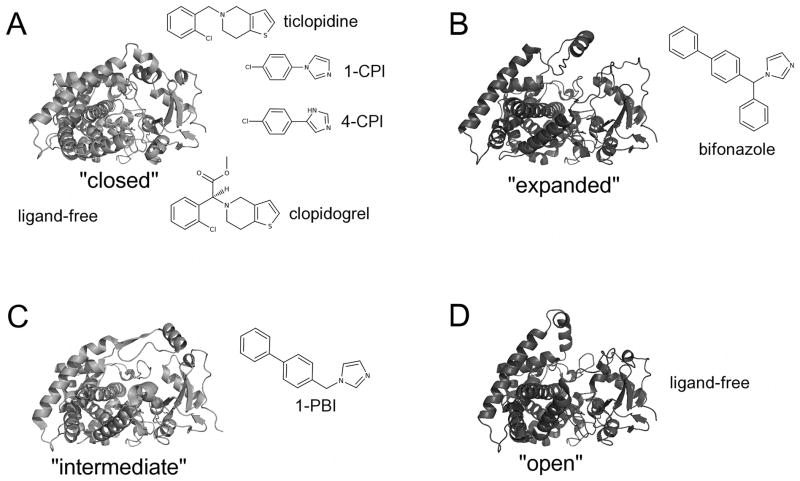
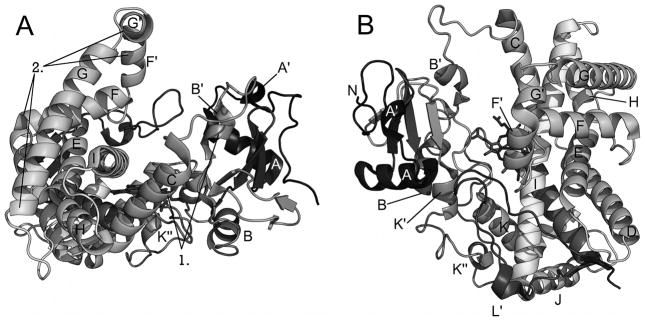
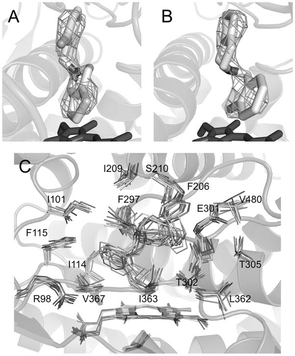
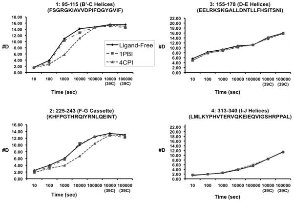
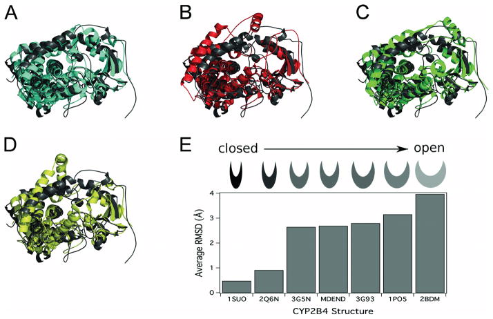
In the past three years, major advances in understanding cytochrome P450 2B (CYP2B) structure-function relationships have been made through determination of multiple ligand-bound and one ligand-free X-ray crystal structure of CYP2B4 and one ligand-bound X-ray crystal structure of CYP2B6. These structures have provided insight into the features that provide the high degree of plasticity of the enzymes. A combination of a phenylalanine cluster that allows for concerted movement of helices F through G and a conserved set of electrostatic interactions involving Arg(262) facilitates movement of this region to accommodate binding of ligands of various sizes without perturbing most of the P450 fold. Integrating solution based techniques such as NMR or deuterium exchange mass spectrometry (DXMS) with computational methods including molecular docking has provided further insight into enzyme behavior upon ligand binding. In addition, extended molecular dynamics simulations have provided a link between an open and a closed conformation of ligand-free CYP2B4 found in crystal structures. Other studies revealed the utility of rational engineering in improving stability of P450s to facilitate structural studies. The solution and computational results combined with the X-ray crystal structures yield a comprehensive picture of how these enzymes adopt different conformations to bind various ligands.


Figures

Figure 1
Figure 1
Conformations observed in CYP2B X-ray crystal structures. CYP2B4 forms a compact structure (A) when bound to the small inhibitors 4-CPI (1SUO), 1-CPI (2Q6N), ticlopidine (3KW4), or clopidogrel (3ME6). Rearrangement of the B′/C loop and F-G cassette accommodates the larger inhibitors (B) bifonazole (2BDM) and (C) 1-PBI (3G5N). Interestingly, the absence of ligand has yielded structures in two conformations: a closed structure similar to the CYP2B4-4-CPI complex (A) and an open conformation (D). Stick diagrams show the chemical structures of ligands.
Figure 2
Figure 2
Structure of CYP2B4. (A) View of CYP2B4 along helix I showing large open cleft with elements of the B′-C loop (1) and F-G cassette (2) labeled. (B) Perspective looking down on heme and active site.
Figure 3
Figure 3
Electron density maps and docking results of CYP2B4 and ticlopidine. The Fo-Fc simulated annealing omit map for ticlopidine contoured at 3σ shows two lobes of electron density above the plane of the heme. Ticlopidine fits this density well either in the thiophene down (A) or the chlorophenyl down (B) orientation. Molecular docking studies using Autodock4 or simulated annealing in GROMACS4 show that the preferred orientation for CYP2B4 binding of ticlopidine is with the chlorophenyl group toward the heme as represented by the results of the simulated annealing experiment (C). Modified from originally published figures in Gay et al. [23] and reproduced with permission from the American Chemical Society. © the American Chemical Society.
Figure 4
Figure 4
CYP2B4 behavior in solution upon ligand binding. Time course of deuterium exchange in the B′-C loop region and the F-G cassette, respectively, of peptides 1 (residues 95–115) and 2 (residues 225–243), which show differences in DXMS exchange. Peptides in the D-E helices and I-J helices, respectively are illustrated as 3 (residues 155–178) and 4 (residues 313–340). The scale of the axis is the maximum number of exchangeable amides. Modified from originally published figure in Wilderman et al. [24] and reproduced with permission from the American Society for Biochemistry and Molecular Biology. © the American Society for Biochemistry and Molecular Biology.
Figure 5
Figure 5
Comparison of results from MD simulation to existing crystal structures of CYP2B4. Overlay of the ending structure of the MD simulation (MDEND, dark gray) with A) the closed ligand-free 2B4dH crystal structure (3MVR, closed conformation, cyan), B) the crystal structure of 2B4dH with bifonazole (2BDM, expanded conformation, red), C) the crystal structure of 2B4dH with three molecules of 1-PBI (3G5N, intermediate conformation, green), or D) the crystal structure of “open” ligand-free 2B4dH (1PO5, open conformation, yellow). E) RMSD of the Cα backbone of the P450 2B4dH crystal structures and MD simulation from the closed ligand-free P450 2B4dH structure (3MVR). The category axis is labeled with the PDB ID of the crystal structure or with MDEND for the average RMSD between 12 ns and 15 ns of the MD simulation (1SUO, P450 2B4dH with 4-CPI; 2Q6N, 2B4dH with 1-CPI; 3G5N, P450 2B4dH with three 1-PBI molecules; 3G93, P450 2B4dH with one 1-PBI molecule; 1PO5, P450 2B4 in the open conformation; 2BDM, P450 2B4dH with bifonazole). Modified from originally published figure in Wilderman et al. [24] and reproduced with permission from the American Society for Biochemistry and Molecular Biology. © the American Society for Biochemistry and Molecular Biology.

Similar articles

Cited by

References

    1. Johnson EF, Stout CD. Structural diversity of human xenobiotic-metabolizing cytochrome P450 monooxygenases. Biochem Bioph Res Co. 2005;338(1):331–336. - PubMed
    1. Wang H, Negishi M. Transcriptional regulation of cytochrome P450 2B genes by nuclear receptors. Curr Drug Metab. 2003;4(6):515–525. - PubMed
    1. Zhao Y, Halpert JR. Structure-function analysis of cytochromes P450 2B. BBA-Gen Subjects. 2007;1770(3):402–412. - PubMed
    1. Lewis DFV, Lake BG, Ito Y, Anzenbacher P. Quantitative structure-activity relationships (QSARS) within cytochromes P450 2B (CYP2B) subfamily enzymes: The importance of lipophilicity for binding and metabolism. Drug Metabol Drug Interact. 2006;21(3–4):213–31. - PubMed
    1. Lewis DFV, Ito Y, Lake BG. Quantitative structure-activity relationships (QSARS) for inhibitors and substrates of CYP2B enzymes: Importance of compound lipophilicity in explanation of potency differences. J Enzym Inhib Med Ch. 2010;25(5):679–684. - PubMed

Publication types

Substances