

Comparative Study
doi: 10.1186/1746-160X-7-25.
Photographic protocol for image acquisition in craniofacial microsomia
Affiliations
- PMID: 22208766
- PMCID: PMC3286411
- DOI: 10.1186/1746-160X-7-25
Item in Clipboard
Comparative Study
Photographic protocol for image acquisition in craniofacial microsomia
Head Face Med.
.
Abstract
Craniofacial microsomia (CFM) is a congenital condition associated with orbital, mandibular, ear, nerve, and soft tissue anomalies. We present a standardized, two-dimensional, digital photographic protocol designed to capture the common craniofacial features associated with CFM.
© 2011 Heike et al; licensee BioMed Central Ltd.
Figures
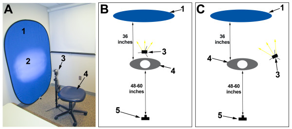
Example of possible configurations for the image environment. Photo of the recommended set up (A) and illustrations of a set up with the monopod flash behind the participant (B) and to the side of the participant (C). (1) Blue background, (2) flash reflection on the background, (3) monopod flash, (4) participant seat, (5) camera.
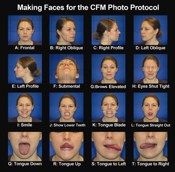
Making Faces for the CFM Photo Protocol. This collage illustrates optimal image acquisition for each of the views described in this protocol. This figure can be used during image acquisition to show participants examples of the requested facial expressions. The grid lines on views A-E can be used on the camera's viewer to ensure optimal head positioning in the Frankfort horizontal plane. The circle overlying images A-E represents the focal point of the image.
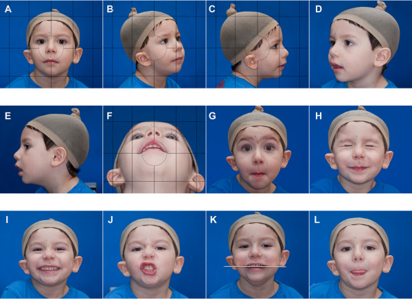
Contact sheet used in the general evaluation in phenotypic assessment tool for CFM. The views obtained during image acquisition can be used to create a contact sheet for quick categorization of the common craniofacial features affected in CFM. The contact sheet illustrated in this figure includes views that can be used to complete the ratings for the orbit, mandible, ear, nerve, and soft tissue in the OMENS classification system. The complete contact sheet incorporates the 16 views obtained in the protocol, in addition to 4 enlarged views of the ears and eyes as illustrated in Figure 4.
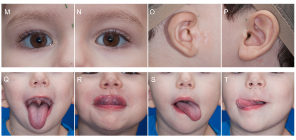
Contact sheet used in the detailed evaluation in phenotypic assessment tool for CFM. This page of the contact sheet is designed to allow raters to quickly assess physical features included in the "Detailed Assessment" of the phenotypic assessment tool for CFM. Enlarged views of the eyes and ears were created by enlarging views A, C, and E. Multiple views of the tongue allow for assessment of unilateral or bilateral hypoplasia.
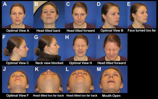
An example of suboptimal images. This collage provides optimal and suboptimal examples of five views. The first three images (A-C) are of View A. Image B is suboptimal for three reasons: the subject's head is tilted back, the blue background is not filling the background completely, and the photographer is angling the camera up for the picture. Image C is suboptimal for two reasons: the subject's head is tilted forward and the photographer is angling the camera down for the picture. The other views represented in this figure include View B (Images D & E), View C (Images F & G), View H (Images H & I), and View F (Images J-M). Image M is suboptimal for two reasons: the subject's mouth is open and the head is not tilted far enough back.
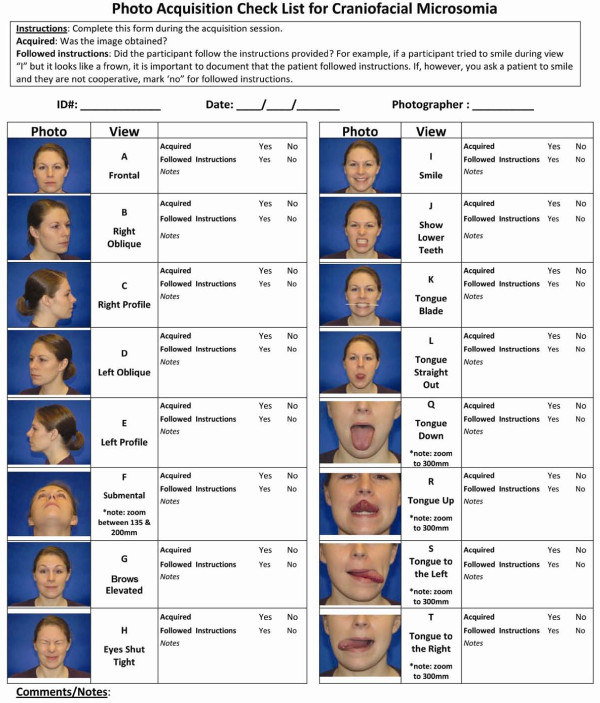
Photo acquisition image check list. This figure provides a check list to be used during image acquisition, along with a section to document notes about the image capture session. These data can be useful for interpretation of the reliability of the images for evaluating nerve function. For example, if the participant tried to smile during the View I, but it looks like a frown, it is important to document that the participant followed instructions. If, however, you ask the participant to smile and they are not cooperative, you would also want to document that as well.
Similar articles
-
Reliable classification of facial phenotypic variation in craniofacial microsomia: a comparison of physical exam and photographs.Head Face Med. 2016 Mar 31;12:14. doi: 10.1186/s13005-016-0109-x. Head Face Med. 2016. PMID: 27029551 Free PMC article.
-
Characterizing facial features in individuals with craniofacial microsomia: A systematic approach for clinical research.Birth Defects Res A Clin Mol Teratol. 2016 Nov;106(11):915-926. doi: 10.1002/bdra.23560. Birth Defects Res A Clin Mol Teratol. 2016. PMID: 27891784 Free PMC article. Clinical Trial.
-
Distinguishing Goldenhar Syndrome from Craniofacial Microsomia.J Craniofac Surg. 2015 Sep;26(6):1887-92. doi: 10.1097/SCS.0000000000002017. J Craniofac Surg. 2015. PMID: 26267577
-
Craniofacial Microsomia.Clin Plast Surg. 2019 Apr;46(2):207-221. doi: 10.1016/j.cps.2018.12.001. Clin Plast Surg. 2019. PMID: 30851752 Review.
-
Distal deletion at 22q11.2 as differential diagnosis in Craniofacial Microsomia: Case report and literature review.Eur J Med Genet. 2018 May;61(5):262-268. doi: 10.1016/j.ejmg.2017.12.013. Epub 2017 Dec 27. Eur J Med Genet. 2018. PMID: 29288792 Review.
Cited by
-
Intelligence and Academic Achievement of Adolescents with Craniofacial Microsomia.Plast Reconstr Surg. 2017 Sep;140(3):571-580. doi: 10.1097/PRS.0000000000003584. Plast Reconstr Surg. 2017. PMID: 28841618 Free PMC article.
-
Interrater reliability of a phenotypic assessment tool for the ear morphology in microtia.Am J Med Genet A. 2013 Jun;161A(6):1264-72. doi: 10.1002/ajmg.a.35963. Epub 2013 Apr 24. Am J Med Genet A. 2013. PMID: 23616389 Free PMC article.
-
Reliable classification of facial phenotypic variation in craniofacial microsomia: a comparison of physical exam and photographs.Head Face Med. 2016 Mar 31;12:14. doi: 10.1186/s13005-016-0109-x. Head Face Med. 2016. PMID: 27029551 Free PMC article.
-
Characterizing facial features in individuals with craniofacial microsomia: A systematic approach for clinical research.Birth Defects Res A Clin Mol Teratol. 2016 Nov;106(11):915-926. doi: 10.1002/bdra.23560. Birth Defects Res A Clin Mol Teratol. 2016. PMID: 27891784 Free PMC article. Clinical Trial.
-
Parental Reports of Intervention Services and Prevalence of Teasing in a Multinational Craniofacial Microsomia Pediatric Study.J Craniofac Surg. 2021 Nov-Dec 01;32(8):2687-2691. doi: 10.1097/SCS.0000000000007999. J Craniofac Surg. 2021. PMID: 34727472 Free PMC article.
References
-
- Heike CL, Hing AV. In: GeneReviews [Internet] Pagon RA, Bird TD, Dolan CR, et al, editor. Seattle (WA): University of Washington, Seattle; 1993. Craniofacial Microsomia Overview. 2009. - PubMed
-
- Gorlin RJ, Cohen MM, Jr, Hennekam RCM. Syndromes of the Head and Neck. Fourth. Oxford University Press; 2001.
Publication types
MeSH terms
Grants and funding
LinkOut - more resources
Full Text Sources
