. 2012:502:241-71.
doi: 10.1016/B978-0-12-416039-2.00013-6.

Oral enzyme therapy for celiac sprue

Affiliations

Oral enzyme therapy for celiac sprue

Michael T Bethune et al. Methods Enzymol. 2012.

Abstract

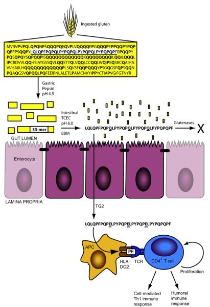
Celiac sprue is an inflammatory disease of the small intestine caused by dietary gluten and treated by adherence to a life-long gluten-free diet. The recent identification of immunodominant gluten peptides, the discovery of their cogent properties, and the elucidation of the mechanisms by which they engender immunopathology in genetically susceptible individuals have advanced our understanding of the molecular pathogenesis of this complex disease, enabling the rational design of new therapeutic strategies. The most clinically advanced of these is oral enzyme therapy, in which enzymes capable of proteolyzing gluten (i.e., glutenases) are delivered to the alimentary tract of a celiac sprue patient to detoxify ingested gluten in situ. In this chapter, we discuss the key challenges for discovery and preclinical development of oral enzyme therapies for celiac sprue. Methods for lead identification, assay development, gram-scale production and formulation, and lead optimization for next-generation proteases are described and critically assessed.


Figures

Figure 1
Figure 1
Gastrointestinal digestion of gluten releases metastable immunogenic peptides. The sequence for α2-gliadin, a representative gluten protein, is shown with proline (17%) and glutamine (36%) content in bold. The 33-mer sequence is underlined. Pepsin cleaves α2-gliadin in the stomach into large peptides, which empty into the upper small intestine. In the intestinal lumen, pancreatic proteases (trypsin, chymotrypsin, elastase, and carboxypeptidase A; TCEC) and intestinal brush border membrane (BBM) peptidases digest most peptides to single amino acids, di-, and tri-peptides for absorption. The 33-mer, however, persists through digestion to traverse the epithelial barrier, becoming deamidated by transglutaminase 2 (TG2) at select glutamine residues (underlined). In the underlying lamina propria, epitopes derived from the deamidated 33-mer (schematized as “PE” to emphasize their proline and deamidated glutamine content) are presented by HLA-DQ2+ antigen-presenting cells (APC), eliciting proliferation and deleterious immune activation from gluten-specific, DQ2-restricted CD4+ T cells. Gastrointestinal glutenases administered during oral enzyme therapy are proposed to digest gluten in the stomach or gut, thereby preventing immune activation.
Figure 2
Figure 2
Features of the immunogenic 33-mer gluten peptide. Prolines (P; 39%) and glutamines (Q; 30%) are over-represented, imparting resistance to gastrointestinal digestion. Transglutaminase 2-mediated deamidation of QXP motifs at underlined glutamines introduces negatively-charged glutamates (E), thereby unmasking six overlapping HLA-DQ2-restricted T cell epitopes (underlined) present in the multivalent 33-mer. Cleavage sites for glutenase candidates EP-B2 (filled arrowheads) and SC PEP (open arrowheads) are indicated.
Figure 3
Figure 3
Chromogenic and fluorogenic substrates. Substrate positions flanking the cleavage site (▼) are indicated as …P2-P1-▼-P1′-P2′… according to standard protease nomenclature (Berger and Schechte.I, 1970).
Figure 4
Figure 4
Detection of 33-mer by liquid chromatography-assisted triple quadrupole mass spectrometry (3Q LC-MS). A) Schematic of the 3Q LC-MS instrument, configured to run in multiple reaction monitoring (MRM) mode. Gluten digests, serum samples, or other complex biological samples are depleted of proteins (>10 kDa) by precipitation with acetonitrile (ACN). Peptides remaining in the supernatant are injected on reversed-phase HPLC and separated according to hydrophobicity. The retention time for 33-mer is 6.4 minutes in the example shown. Peptides eluted from the column are ionized by electrospray ionization and precursor ions enter the first quadrupole. Undesired ion species are filtered out, whereas [33-mer+3H]3+ is selected for fragmentation by collision with an inert gas in the collision chamber. Fragment ions then pass through the third quadrupole, which again filters out undesired species and transmits only those selected ions to the detector. B) Spectra showing the precursor ion selected in the first quadrupole (left) and the fragment ions selected in the third quadrupole (right). Note that another precursor ion, [33-mer+4H]4+ is prominent in the first quadrupole. This precursor ion can also be selected for fragmentation in MRM mode, providing additional precursor → fragment transitions. Chromatograms (bottom) show peaks at the expected retention time for the two transitions monitored, as well as for the total ion current. The analyte is quantified by integrating an individual transition or the TIC chromatogram peak, normalizing to an internal control (e.g. isotope-labeled 33-mer), and comparing normalized area-under-the-curve to a standard curve.
Figure 5
Figure 5
Detoxification of gluten from whole-wheat bread using an acid-active two-enzyme glutenase. Whole-wheat bread was digested with pepsin supplemented with varied concentrations of EP-B2 ± SC PEP for 60 min at pH 4.5 at 37 °C. A. Reversed-phase HPLC analysis of effect of EP-B2 ± SC PEP on gluten digestion. The broad peak between 12.5-22 minutes reflects immunogenic gluten peptides 9-22 residues in length. Note that ordinate axis is scaled to highlight the incremental effect of increasing SC PEP; this view truncates the TAME internal standard peak. B. T cell proliferation assays measuring antigen content of whole-wheat bread digests. Pepsin (0.6 mg/mL) was supplemented with EP-B2 (200 U) ± SC PEP (0.5 U). Gastric digests were treated for an additional 10 min under duodenal conditions (0.375 mg/mL trypsin and chymotrypsin and 0.075 mg/mL elastase and carboxypeptidase A at pH 6.0 at 37 °C) to solubilize all remaining T cell epitopes. P28 and P35 are T cell lines expanded from celiac patient intestinal biopsies. A stimulation index of 1 indicates background levels of proliferation.

References

    1. Alaedini A, Green PH. Narrative review: celiac disease: understanding a complex autoimmune disorder. Ann. Intern. Med. 2005;142:289–98. - PubMed
    1. Bethune MT, Crespo-Bosque M, Bergseng E, Mazumdar K, Doyle L, Sestak K, Sollid LM, Khosla C. Noninflammatory gluten peptide analogs as biomarkers for celiac sprue. Chem Biol. 2009;16:868–81. - PMC - PubMed
    1. Bethune MT, Khosla C. Parallels between pathogens and gluten peptides in celiac sprue. PLoS Pathog. 2008;4:e34. - PMC - PubMed
    1. Bethune MT, Ribka E, Khosla C, Sestak K. Transepithelial transport and enzymatic detoxification of gluten in gluten-sensitive rhesus macaques. PLoS ONE. 2008;3:e1857. - PMC - PubMed
    1. Bethune MT, Strop P, Tang Y, Sollid LM, Khosla C. Heterologous expression, purification, refolding, and structural-functional characterization of EP-B2, a self-activating barley cysteine endoprotease. Chem. Biol. 2006;13:637–47. - PubMed

MeSH terms