. 2012 Jan 17;22(1):38-51.
doi: 10.1016/j.devcel.2011.11.013. Epub 2011 Dec 29.

DUX4 activates germline genes, retroelements, and immune mediators: implications for facioscapulohumeral dystrophy

Affiliations

DUX4 activates germline genes, retroelements, and immune mediators: implications for facioscapulohumeral dystrophy

Linda N Geng et al. Dev Cell. .

Abstract

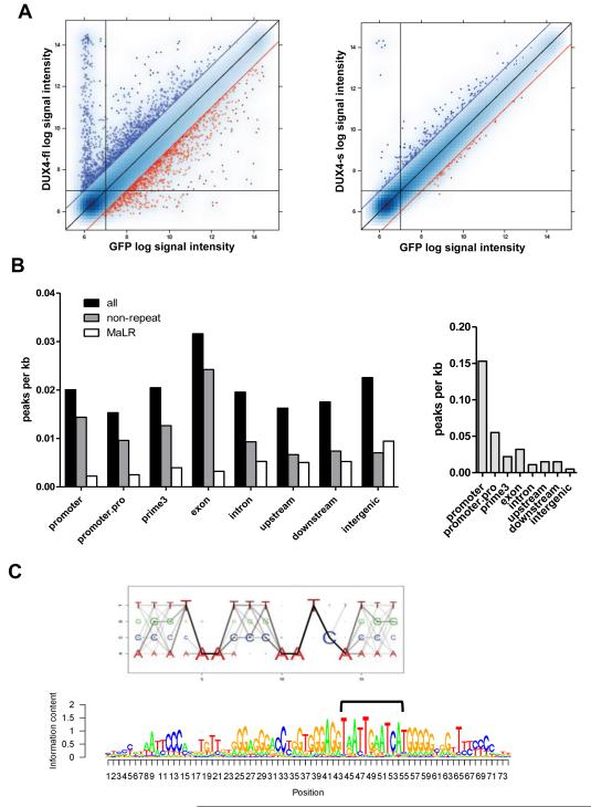
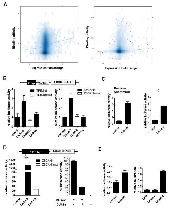
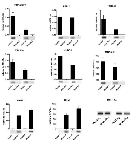
Facioscapulohumeral dystrophy (FSHD) is one of the most common inherited muscular dystrophies. The causative gene remains controversial and the mechanism of pathophysiology unknown. Here we identify genes associated with germline and early stem cell development as targets of the DUX4 transcription factor, a leading candidate gene for FSHD. The genes regulated by DUX4 are reliably detected in FSHD muscle but not in controls, providing direct support for the model that misexpression of DUX4 is a causal factor for FSHD. Additionally, we show that DUX4 binds and activates LTR elements from a class of MaLR endogenous primate retrotransposons and suppresses the innate immune response to viral infection, at least in part through the activation of DEFB103, a human defensin that can inhibit muscle differentiation. These findings suggest specific mechanisms of FSHD pathology and identify candidate biomarkers for disease diagnosis and progression.


Figures

Figure 1
Figure 1. DUX4-fl activates the expression of germline genes and binds a double-homeobox motif
(A) Pairwise comparison of normalized array data from DUX4-fl vs GFP (left), and DUX4-s vs GFP (right). Blue dots: upregulated genes; red dots: downregulated genes; blue and red diagonal lines represent 2-fold change. Vertical and horizontal lines represent signal thresholds for calling genes present or absent. (B) Gene context of DUX4-fl binding sites (left) or MyoD binding sites (right) represented by peak density, adjusting for prevalence of gene context category in the genome. Promoter: +/−500 bp from the transcription start site (TSS); promoter.pro: +/−2 kb from the TSS; prime3: +/−500 nt from the end of the transcript; upstream: −2 kb to −10 kb upstream of the TSS; downstream: +2 kb to +10 kb from the end of the transcript; intergenic: >10 kb from any annotated gene. (C) Top: DUX4-fl motif Logo. The size of each nucleotide at a given position is proportional to the frequency of the nucleotide at that position, and the darkness of the line connecting two adjacent nucleotides represents corresponding dinucleotide frequency. Bottom: DUX4 binding motif matches MaLR repeat consensus sequence. We identified the best DUX4 binding sites (bracket) within the MaLR repeats annotated in the RepeatMasker track provided by the UCSC genome browser (hg18) and extended the motif in the flanking regions to reflect general MaLR repeat consensus.
Figure 2
Figure 2. DUX4-fl activates transcription in vivo and DUX4-s can interfere with its activity
(A) Comparison of DUX4 binding and regional gene transcription. The DUX4 peak height (Y-axis, square root transformation) of binding sites located within the CTCF-flanked domain of the TSS (left panel) or within +/− 2 KB of the TSS (right panel) plotted against the log-2 fold change of mRNA expression for the gene associated with the TSS (X-axis). The trend line shows a weak association between DUX4 binding and regional gene transcription. (B) Genomic fragments near theTRIM48 and ZSCAN4 genes containing DUX4 binding sites were cloned into pGL3-promoter reporter vector (schematic, top) and transfected into human rhabdomyoscaroma cell line RD. Cells were co-transfected with DUX4-fl or DUX4-s. pCS2-β galactosidase (beta-gal) was used to balance DNA amount in control condition. TRIM48mut and ZSCAN4mut, mutated binding sites. (C) DUX4-fl can act as an enhancer. Left: ZSCAN4 DUX4 binding site in reverse orientation relative to panel B upstream of the SV40 promoter. Right: ZSCAN4 DUX4 binding site in original orientation but moved 3′ downstream of the reporter gene. Luciferase activity set relative to control and error bars represent standard deviation of triplicates. (D) Left panel: Genomic fragment upstream of the ZSCAN4 translation start site containing four DUX4 binding sites was cloned into pGL3-basic luciferase vector (schematic, top). DUX4-fl highly activates the luciferase expression, whereas mutation of the binding sites (ZSCAN4mut) drastically reduces this induction. Right panel: Luciferase activity from DUX4-fl is set at 100% (pCS2-beta-gal is used to normalize the amount of plasmid). Co-transfection of equal amounts of DUX4-fl and DUX4-s diminishes luciferase activity. (E) Left: Genomic fragment from the LTR of THE1D MaLR element containing the DUX4 binding site were cloned into pGL3-promoter vector and tested for response to DUX4-fl as in (a). Right: Transcripts from endogenous retroelement MaLRs are upregulated by lentiviral transduction of DUX4-fl into primary human myoblasts. No upregulation is seen with lentiviral transduction of GFP or DUX4-s. Real-time RT-PCR quantitation is reported relative to internal standard RPL13a. All data represent mean +/− SD from at least triplicates.
Figure 3
Figure 3. DUX4 targets are normally expressed in human testis but not in healthy skeletal muscle
DUX4-fl targets expression in human testis versus matched skeletal muscle tissue from two healthy donors. Real-time RT-qPCR analysis of gene expression is presented relative to internal standard RPL13a and error bars represent standard deviation of PCR triplicates. MYH2 and CKM are markers of skeletal muscle. Qualitative RT-PCR gel panels are shown below qPCR graphs.
Figure 4
Figure 4. DUX4 regulated genes normally expressed in the testis are aberrantly expressed in FSHD muscle
Real-time RT-PCR quantitation of six DUX4-fl target genes, PRAMEF1, RFPL2, TRIM43, ZSCAN4, KHDC1 and MBD3L2. Values are expressed as relative to internal standard RPL13a and represent mean +/− SD from triplicates. See Table S5 for all sample names and endogenous DUX4-fl expression status. (A) Cultured control and FSHD muscle cells. Note, sample 11 is from FSHD2 individual. (B) Control and FSHD muscle biopsies. (C) Top: RT-PCR gel showing siRNA knockdown of endogenous DUX4-fl in cultured FSHD muscle cells, done in triplicate with Timm17b as an internal standard. Negative control siRNA is against unrelated luciferase gene. Bottom: Levels of DUX4-fl target genes relative to the control treated samples were also reduced, as measured by qPCR. Error bars represent SD of triplicates; *P<0.05, **P<0.01 between DUX4 siRNA and control siRNA treated cells.
Figure 5
Figure 5. DEFB103 inhibits innate immune response to viral infection and inhibits muscle differentiation
Real time RT-PCR quantitation of innate immune responsive genes and genes involved in muscle differentiation. Values represent mean +/− SD from triplicates and are either expressed as relative to internal standard RPL13a or as percentage relative to control condition after being normalized to RPL13a. (A) Expression levels of innate immune responder IFIH1 and secreted factor DEFB103 after infection of muscle cells with lenti-GFP or lenti-DUX4-fl. (B) Expression levels of innate immune responders IFIH1 and ISG20 after infection with lenti-GFP in the presence of untreated media (control) or media supplemented with human β-defensin 3 peptide (5.0 ug/ml) (DEFB103) or conditioned media from lenti-DUX4-fl (C.M.). (C) Endogenous expression of DEFB103 in control testis and skeletal muscle tissues (Testis 1/2, Muscle 1/2), FSHD muscle biopsies (F-Muscle 3/4) and cultured FSHD and control muscle cells (F-MB and MB). (D) Upregulation of myostatin (MSTN) in myoblasts cultured in growth media supplemented with 2.5 ug/ml DEFB103. (E) Expression levels of various muscle marker genes in response to 2.5 ug/ml uM DEFB103 when added to muscle cells cultured in differentiation media. Myf6 and Desmin were included as genes that were unchanged on the arrays. (F) Cultured human muscle cells in differentiation media for 72 hr either without (a and b) or with (c and d) 2.5 μg/ml DEFB103. a and c, phase contrast showing inhibition of fusion by DEFB103. b and d, myosin heavy chain immunofluorescence (red) and DAPI nuclear stain (blue).

References

    1. Belancio VP, Roy-Engel AM, Deininger PL. All y’all need to know ‘bout retroelements in cancer. Semin Cancer Biol. 2010;20:200–210. - PMC - PubMed
    1. Bonnefont J, Nikolaev SI, Perrier AL, Guo S, Cartier L, Sorce S, Laforge T, Aubry L, Khaitovich P, Peschanski M, et al. Evolutionary forces shape the human RFPL1,2,3 genes toward a role in neocortex development. Am J Hum Genet. 2008;83:208–218. - PMC - PubMed
    1. Booth HA, Holland PW. Annotation, nomenclature and evolution of four novel homeobox genes expressed in the human germ line. Gene. 2007;387:7–14. - PubMed
    1. Bosch N, Caceres M, Cardone MF, Carreras A, Ballana E, Rocchi M, Armengol L, Estivill X. Characterization and evolution of the novel gene family FAM90A in primates originated by multiple duplication and rearrangement events. Hum Mol Genet. 2007;16:2572–2582. - PubMed
    1. Bosnakovski D, Xu Z, Gang EJ, Galindo CL, Liu M, Simsek T, Garner HR, Agha-Mohammadi S, Tassin A, Coppee F, et al. An isogenetic myoblast expression screen identifies DUX4-mediated FSHD-associated molecular pathologies. Embo J. 2008;27:2766–2779. - PMC - PubMed

Publication types

MeSH terms