. 2012 Feb 7;22(3):180-90.
doi: 10.1016/j.cub.2011.12.029. Epub 2011 Dec 29.

Mad2 and Mad3 cooperate to arrest budding yeast in mitosis

Affiliations

Mad2 and Mad3 cooperate to arrest budding yeast in mitosis

Derek T C Lau et al. Curr Biol. .

Abstract

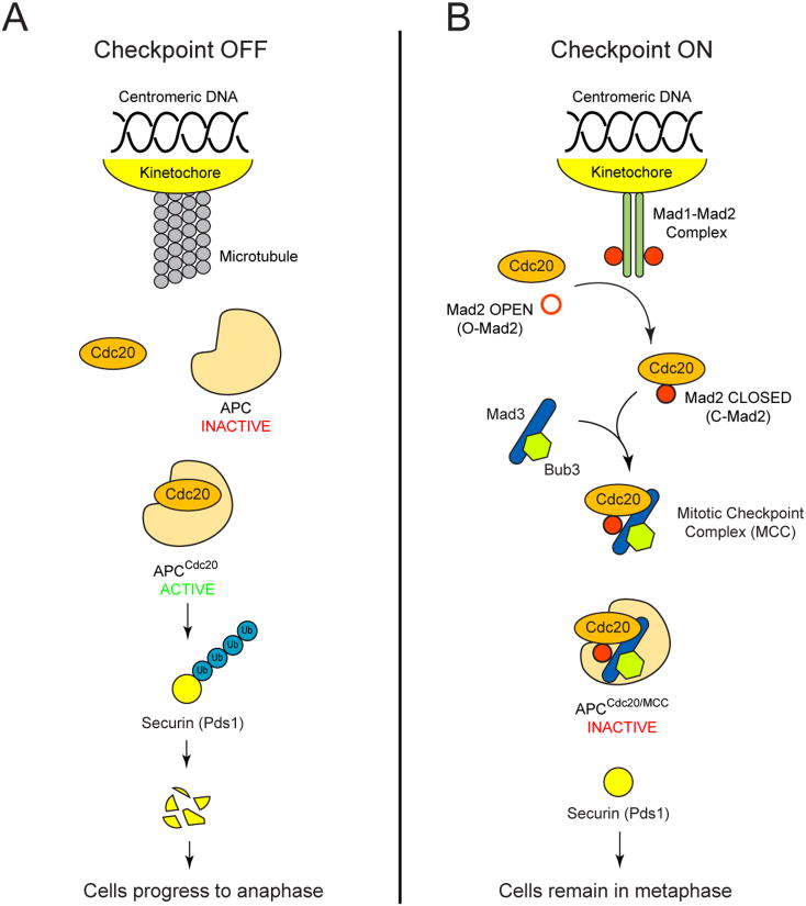
Background: The spindle checkpoint ensures accurate chromosome transmission by delaying chromosome segregation until all chromosomes are correctly aligned on the mitotic spindle. The checkpoint is activated by kinetochores that are not attached to microtubules or are attached but not under tension and arrests cells at metaphase by inhibiting the anaphase-promoting complex (APC) and its coactivator Cdc20. Despite numerous studies, we still do not understand how the checkpoint proteins coordinate with each other to inhibit APC(Cdc20) activity.

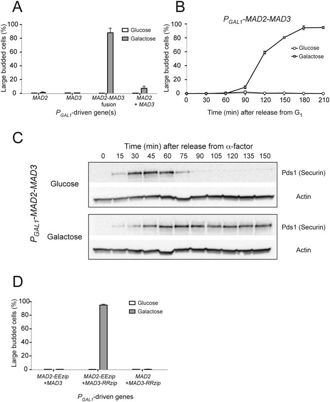
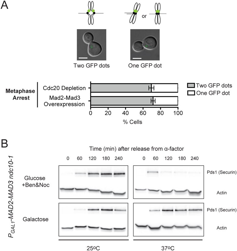
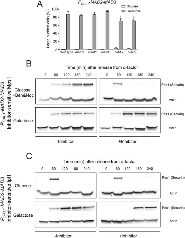
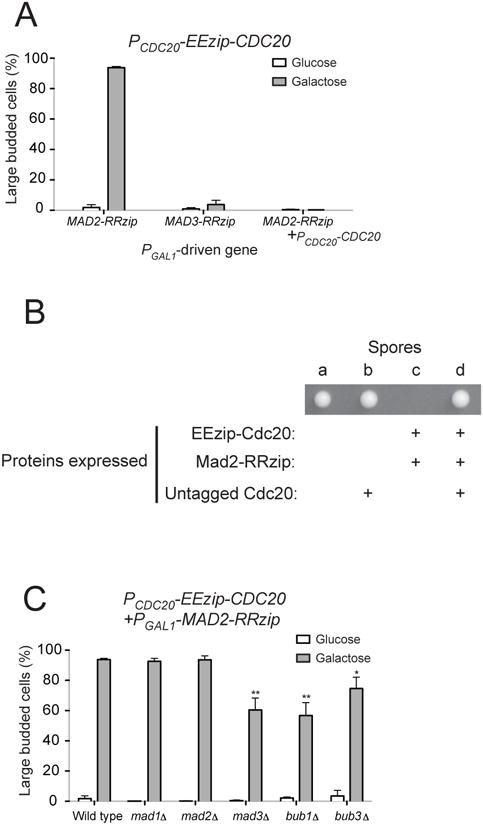
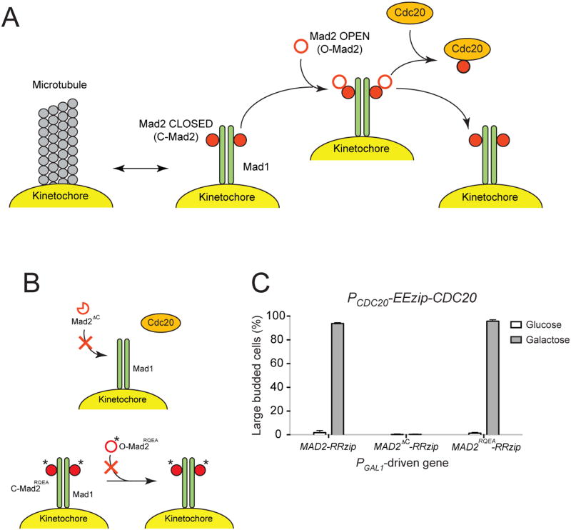
Results: To ask how the checkpoint components induce metaphase arrest, we constructed fusions of checkpoint proteins and expressed them in the budding yeast Saccharomyces cerevisiae to mimic possible protein interactions during checkpoint activation. We found that expression of a Mad2-Mad3 protein fusion or noncovalently linked Mad2 and Mad3, but not the overexpression of the two separate proteins, induces metaphase arrest that is independent of functional kinetochores or other checkpoint proteins. We further showed that artificially tethering Mad2 to Cdc20 also arrests cells in metaphase independently of other checkpoint components.

Conclusion: Our results suggest that Mad3 is required for the stable binding of Mad2 to Cdc20 in vivo, which is sufficient to inhibit APC activity and is the most downstream event in spindle checkpoint activation.


Figures

Figure 1
Figure 1
A model for spindle checkpoint activation (adapted from [6, 7]). (A) During mitosis, when all chromosomes are properly attached to microtubules, the anaphase-promoting complex (APC) and its co-activator Cdc20 polyubiquitinate different substrates such as securin (Pds1 in budding yeast), leading to its destruction and anaphase onset. (B) The spindle checkpoint is activated by the absence of microtubule attachment or the lack of tension at the kinetochore. Mad1-Mad2 complexes associate with unattached kinetochores and recruit the “open” Mad2 conformer (O-Mad2), facilitating the formation of a “closed” Mad2 (C-Mad2)-Cdc20 complex. The closed Mad2-Cdc20 complex associates with Mad3 and Bub3, which can be recruited to the kinetochores by interactions with Bub1 (not shown), to form the mitotic checkpoint complex (MCC). Inhibition of APC activity by the MCC arrests the cells in metaphase and gives the cells time to correct attachment errors at the kinetochores.
Figure 2
Figure 2
Overexpressing a Mad2-Mad3 protein fusion leads to metaphase arrest. (A) Cells with the indicated PGAL1-driven genes were grown to mid-log phase, arrested in G1 with α-factor, and were released into media with either glucose or galactose. After 3 hours of growth, the percentage of large budded cells was determined by light microscopy as a measure of metaphase arrest. Error bars represent the standard deviation of three independent trials. Two hundred cells were counted for each trial. (B) PGAL1-MAD2-MAD3 cells were released from G1 arrest into glucose- or galactose-containing media. Samples were collected at the indicated time point and the percentage of large budded cells was determined by light microscopy. Error bars represent the standard deviation of three independent trials. Two hundred cells were counted at each time point for each trial. (C) Cell cycle progression of PGAL1-MAD2-MAD3 cells was monitored by Western blots (n=3), which detect securin (Pds1), a protein that is destroyed as cells enter anaphase. Cells were grown to mid-log phase and arrested in G1 with α-factor, and were released into media with either glucose (top) or galactose (bottom). Western blots against Myc (to visualize Myc-tagged securin) or actin (loading control) were performed. When the cells were grown in glucose, securin level first increased and then dropped rapidly as cells progressed into anaphase. When the Mad2-Mad3 fusion was overexpressed in the presence of galactose, securin was stabilized, indicating that the cells were arrested in metaphase. (D) Overexpressing Mad2 and Mad3 linked by leucine zippers also induces metaphase arrest. Cells with the indicated PGAL1-driven genes were released from G1 arrest into glucose- or galactose-containing media. The percentage of large budded cells was determined by light microscopy after 3 hours of growth. Error bars represent the standard deviation of three independent trials. Two hundred cells were counted for each trial.
Figure 3
Figure 3
Metaphase arrest by the Mad2-Mad3 fusion is independent of kinetochore-microtubule attachments. (A) Chromosomes bi-orient in cells overexpressing the Mad2-Mad3 fusion. To achieve metaphase arrest, cells carrying PGAL1-MAD2-MAD3, GFP-LacI, a LacO array located near the centromere of chromosome XV, and PMET3-CDC20 were released from G1 arrest into either medium with glucose and methionine (Cdc20 depletion) or medium with galactose and no methionine (Mad2-Mad3 overexpression). Differential interference contrast (DIC) and GFP images of the cells were taken 3 hours after their release from G1. One or two GFP dots can be seen in cells arrested in metaphase. Cells have two GFP dots when chromosome XV bi-orients and the two sister kinetochores are separated by the spindle. Cells have one GFP dot when chromosome XV mono-orients (sister kinetochores attaching to the same spindle pole) or bi-orients but the two sister kinetochores are not pulled apart enough to allow resolution of two separate dots. Representative images of metaphase-arrested cells with one or two GFP dots are shown (Scale bar, 5 μm). Bar graph shows the percentage of cells with one or two GFP dots when they were arrested by Cdc20 depletion or overexpression of the Mad2-Mad3 fusion. Error bars represent the standard deviation of three independent trials. Two hundred cells were counted for each trial. Around 70% of cells arrested by Cdc20 depletion had two GFP dots. The percentage of cells that showed two GFP dots when the cells were arrested by the Mad2-Mad3 fusion is statistically indistinguishable (P=0.415; two-tailed Student's t test). Cells therefore show normal chromosome bi-orientation when they are arrested in metaphase by the Mad2-Ma3 fusion, suggesting that the fusion does not disrupt kinetochore-microtubule connections. (B) Metaphase arrest by the Mad2-Mad3 fusions does not require functional kinetochores. Cell cycle progression of cells with PGAL1-MAD2-MAD3 and ndc10-1 (a mutation that inactivates kinetochore at 37°C) was measured by Western blots (n=3). Cells were released from G1 arrest into media with either glucose with benomyl and nocodazole (top) or galactose (bottom) at 25°C or 37°C. Western blots against Myc (to visualize Myc-tagged securin) or actin (loading control) were performed. Securin was stabilized in galactose-containing medium even at 37°C, indicating that the metaphase arrest is independent of functional kinetochores.
Figure 4
Figure 4
Metaphase arrest by Mad2-Mad3 fusions does not require other checkpoint components. (A) Cells with PGAL1-MAD2-MAD3 and deletion of the indicated checkpoint genes were released from G1 arrest into glucose- or galactose-containing media. The percentage of large budded cells was determined by light microscopy after 3 hours of growth. Error bars represent the standard deviation of three independent trials. Two hundred cells were counted for each trial. Asterisks indicate significant difference from wild-type control (*P < 0.05; two-tailed Student's t test). (B) Metaphase arrest by the Mad2-Mad3 fusion does not require Mps1 function. Cell cycle progression of cells with PGAL1-MAD2-MAD3 and mps1-as1 was monitored by Western blotting (n=3). Cells were released from G1 arrest into media with either glucose with benomyl and nocodazole (top) or galactose (bottom), in the absence (-Inhibitor) or presence (+Inhibitor) of 1NM-PP1, an inhibitor of the analog-sensitive Mps1. Western blots against Myc or actin (loading control) were performed. Both securin and Mps1-as1 are tagged with Myc in the strain, but only the bands corresponding to Myc-tagged securin are shown in the figure. Securin was stabilized in galactose-containing medium even in the presence of inhibitor, indicating that Mps1 activity is not needed for the metaphase arrest. The cause of the delay in accumulation of securin in cells grown in medium with galactose and the inhibitor is not known. (C) The Mad2-Mad3 fusion does not require Ipl1 activity to induce metaphase arrest. The cell cycle progression of cells with PGAL1-MAD2-MAD3 and ipl1-as5 was measured by Western blots (n=3). Cells were released from G1 arrest into media with either glucose (top) or galactose (bottom), in the absence (-Inhibitor) or presence (+Inhibitor) of 1NA-PP1, an inhibitor of the analog-sensitive Ipl1. Western blots against Myc (to visualize Myc-tagged securin) or actin (loading control) were performed. Securin was stabilized in galactose-containing medium in the presence of inhibitor, showing that Ipl1 activity is not needed for the metaphase arrest. The cause of the delay in accumulation of securin in cells grown in medium with galactose and the inhibitor is not known.
Figure 5
Figure 5
Tethering Mad2 directly to Cdc20 leads to metaphase arrest. (A) Cells with PCDC20-EEzip-CDC20 and the indicated PGAL1-driven genes were released from G1 arrest into glucose- or galactose-containing media. The percentage of large budded cells was determined by light microscopy after 3 hours of growth. Error bars represent the standard deviation of three independent trials. Two hundred cells were counted for each trial. Cells in the last column contain PCDC20-EEzip-CDC20, PGAL1-MAD2-RRzip, and an untagged copy of Cdc20 (PCDC20-CDC20) and thus cannot be arrested in mitosis by expressing Mad2-RRzip. (B) Cells expressing EEzip-Cdc20 and Mad2-RRzip from the endogenous CDC20 and MAD2 promoters display growth defects that can be overcome by expressing untagged Cdc20. Diploids that are heterozygous for three manipulated genes, PCDC20-EEzip-CDC20, PMAD2-MAD2-RRzip, and PCDC20-CDC20@URA3, were sporulated and a total of 15 tetrads were dissected (also see Table 1). Representative image of a tetrad on rich, glucose-containing plate after 2 days of growth at 30°C is shown. The “+” signs indicate proteins that are expressed based on the genotypes of each spore, which were determined by replica plating the tetrad onto dropout or drug plates. The genotype of spore c, which failed to form visible colonies, was inferred from the genotypes of other spores from the same tetrad. (C) The metaphase arrest produced by tethering Mad2 to Cdc20 does not require other checkpoint components (also see Figure S1). Cells with PCDC20-EEzip-CDC20, PGAL1-MAD2-RRzip, and deletion of the indicated checkpoint genes were released from G1 arrest into glucose-or galactose-containing media. The percentage of large budded cells was determined by light microscopy after 3 hours of growth. Error bars represent the standard deviation of three independent trials. Two hundred cells were counted for each trial. Asterisks indicate significant difference from wild-type control (*P < 0.05 and **P < 0.01; two-tailed Student's t test).
Figure 6
Figure 6
. Metaphase arrest induced by tethering Mad2 mutants to Cdc20 supports the Mad2-template model. (A) The Mad2-template model (adapted from [6]). Mad1 dimers associate with unattached kinetochores and bind Mad2, converting them from “open” (O-Mad2) to “closed” (C-Mad2) conformation. The Mad1-Mad2 complexes at the kinetochores (the “templates”) then recruit additional open Mad2, allowing the formation of closed Mad2-Cdc20 complexes. (B) The behavior of Mad2 mutants in the context of the Mad2-template model. (Top) Mad2ΔC lacks the C-terminal amino acid residues and cannot convert to the closed Mad2 conformation. It fails to activate the spindle checkpoint since it is unable to form a stable complex with Mad1 and to bind to and inhibit Cdc20. (Bottom) The double point mutant Mad2RQEA carries the mutations Arg126-Glu and Gln127-Ala. The changes inhibit the binding between free Mad2 and closed conformation of Mad2 found in the Mad1-Mad2 complex, which inactivates the spindle checkpoint by preventing the formation of C-Mad2-Cdc20 complexes. The mutations also affect the interaction of Mad2 with BUBR1 (mammalian version of Mad3) and the formation of stable MCC. (C) Effects of tethering Mad2 mutants to Cdc20. Cells with PCDC20-EEzip-CDC20 and the indicated PGAL1-driven genes were released from G1 arrest into glucose- or galactose-containing media. The percentage of large budded cells was determined by light microscopy after 3 hours of growth. Error bars represent the standard deviation of three independent trials. Two hundred cells were counted for each trial. Tethering the Mad2 mutant that can reach the closed conformation, but cannot induced conformational conversion in other Mad2 molecules (Mad2RQEA), does activate the checkpoint, but tethering the mutant that cannot achieve the closed conformation (Mad2ΔC) does not.

Comment in

Similar articles

Cited by

References

    1. Li R, Murray AW. Feedback control of mitosis in budding yeast. Cell. 1991;66:519–531. - PubMed
    1. Hoyt MA, Totis L, Roberts BT. S. cerevisiae genes required for cell cycle arrest in response to loss of microtubule function. Cell. 1991;66:507–517. - PubMed
    1. Li X, Nicklas RB. Mitotic forces control a cell-cycle checkpoint. Nature. 1995;373:630–632. - PubMed
    1. Stern BM, Murray AW. Lack of tension at kinetochores activates the spindle checkpoint in budding yeast. Curr Biol. 2001;11:1462–1467. - PubMed
    1. Biggins S, Murray AW. The budding yeast protein kinase Ipl1/Aurora allows the absence of tension to activate the spindle checkpoint. Genes and Development. 2001;15:3118–3129. - PMC - PubMed

Publication types

MeSH terms