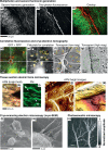
Imaging cardiac extracellular matrices: a blueprint for regeneration
- PMID: 22209562
- PMCID: PMC4139096
- DOI: 10.1016/j.tibtech.2011.12.001
Imaging cardiac extracellular matrices: a blueprint for regeneration
Abstract
Once damaged, cardiac tissue does not readily repair and is therefore a primary target of regenerative therapies. One regenerative approach is the development of scaffolds that functionally mimic the cardiac extracellular matrix (ECM) to deliver stem cells or cardiac precursor populations to the heart. Technological advances in micro/nanotechnology, stem cell biology, biomaterials and tissue decellularization have propelled this promising approach forward. Surprisingly, technological advances in optical imaging methods have not been fully utilized in the field of cardiac regeneration. Here, we describe and provide examples to demonstrate how advanced imaging techniques could revolutionize how ECM-mimicking cardiac tissues are informed and evaluated.
Copyright © 2012 Elsevier Ltd. All rights reserved.
Figures

Similar articles
-
Extracellular Matrix from Whole Porcine Heart Decellularization for Cardiac Tissue Engineering.Methods Mol Biol. 2018;1577:95-102. doi: 10.1007/7651_2017_31. Methods Mol Biol. 2018. PMID: 28456953
-
Preparation of cardiac extracellular matrix scaffolds by decellularization of human myocardium.J Biomed Mater Res A. 2014 Sep;102(9):3263-72. doi: 10.1002/jbma.35000. J Biomed Mater Res A. 2014. PMID: 24142588
-
Extracellular Matrix for Myocardial Repair.Adv Exp Med Biol. 2018;1098:151-171. doi: 10.1007/978-3-319-97421-7_8. Adv Exp Med Biol. 2018. PMID: 30238370 Review.
-
Cells, Scaffolds and Their Interactions in Myocardial Tissue Regeneration.J Cell Biochem. 2017 Aug;118(8):2454-2462. doi: 10.1002/jcb.25912. Epub 2017 Apr 25. J Cell Biochem. 2017. PMID: 28128477 Review.
-
Decellularized zebrafish cardiac extracellular matrix induces mammalian heart regeneration.Sci Adv. 2016 Nov 18;2(11):e1600844. doi: 10.1126/sciadv.1600844. eCollection 2016 Nov. Sci Adv. 2016. PMID: 28138518 Free PMC article.
Cited by
-
3D bioprinting for cardiovascular regeneration and pharmacology.Adv Drug Deliv Rev. 2018 Jul;132:252-269. doi: 10.1016/j.addr.2018.07.014. Epub 2018 Jul 24. Adv Drug Deliv Rev. 2018. PMID: 30053441 Free PMC article. Review.
-
Solid organ fabrication: comparison of decellularization to 3D bioprinting.Biomater Res. 2016 Aug 31;20(1):27. doi: 10.1186/s40824-016-0074-2. eCollection 2016. Biomater Res. 2016. PMID: 27583168 Free PMC article. Review.
-
Harnessing native blueprints for designing bioinks to bioprint functional cardiac tissue.iScience. 2025 Jan 23;28(3):111882. doi: 10.1016/j.isci.2025.111882. eCollection 2025 Mar 21. iScience. 2025. PMID: 40177403 Free PMC article. Review.
-
Laminin 411 mediates endothelial specification via multiple signaling axes that converge on β-catenin.Stem Cell Reports. 2022 Mar 8;17(3):569-583. doi: 10.1016/j.stemcr.2022.01.005. Epub 2022 Feb 3. Stem Cell Reports. 2022. PMID: 35120622 Free PMC article.
-
Single-cell analysis of embryoid body heterogeneity using microfluidic trapping array.Biomed Microdevices. 2014 Feb;16(1):79-90. doi: 10.1007/s10544-013-9807-3. Biomed Microdevices. 2014. PMID: 24085533 Free PMC article.
References
-
- Akhyari P, et al. Myocardial tissue engineering: the extracellular matrix. Eur. J. Cardiothorac. Surg. 2008;34:229–241. - PubMed
Publication types
MeSH terms
Grants and funding
LinkOut - more resources
Full Text Sources

