Review
. 2012 Jun 15;71(12):1081-9.
doi: 10.1016/j.biopsych.2011.11.012. Epub 2011 Dec 29.

Optogenetics, sex, and violence in the brain: implications for psychiatry

Affiliations
Review

Optogenetics, sex, and violence in the brain: implications for psychiatry

David J Anderson. Biol Psychiatry. .

Abstract

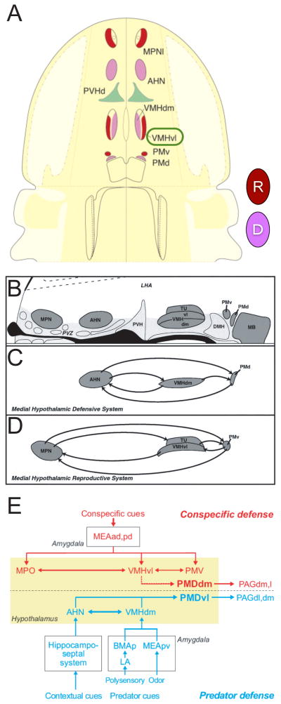
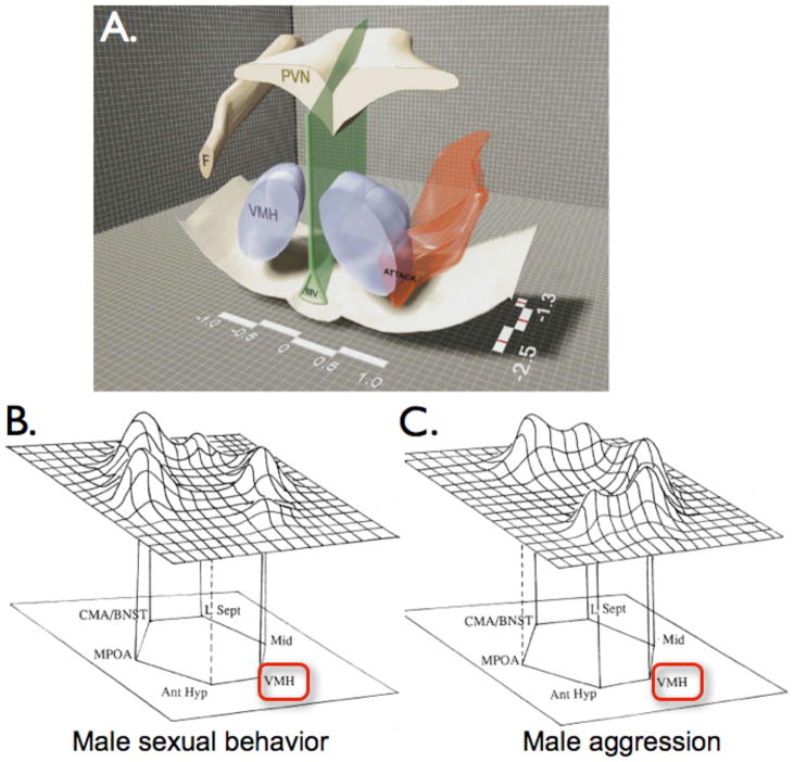
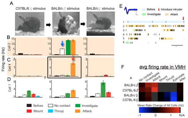
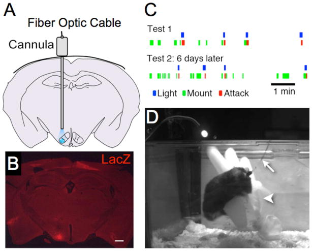
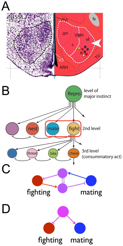
Pathological aggression and the inability to control aggressive impulses takes a tremendous toll on society. Yet aggression is a normal component of the innate behavior repertoire of most vertebrate animal species as well as of many invertebrates. Progress in understanding the etiology of disorders of aggressive behavior, whether genetic or environmental in nature, therefore requires an understanding of the brain circuitry that controls normal aggression. Efforts to understand this circuitry at the level of specific neuronal populations have been constrained by the limited resolution of classical methodologies, such as electrical stimulation and electrolytic lesion. The availability of new, genetically based tools for mapping and manipulating neural circuits at the level of specific, genetically defined neuronal subtypes provides an opportunity to investigate the functional organization of aggression circuitry with cellular resolution. However, these technologies are optimally applied in the mouse, where there has been surprisingly little traditional work on the functional neuroanatomy of aggression. Here we discuss recent, initial efforts to apply optogenetics and other state-of-the-art methods to the dissection of aggression circuitry in the mouse. We find, surprisingly, that neurons necessary and sufficient for inter-male aggression are located within the ventrolateral subdivision of the ventromedial hypothalamic nucleus, a structure traditionally associated with reproductive behavior. These neurons are intermingled with neurons activated during male-female mating, with approximately 20% overlap between the populations. We discuss the significance of these findings with respect to neuroethological and neuroanatomical perspectives on the functional organization of innate behaviors and their potential implications for psychiatry.


Conflict of interest statement

The author reports no biomedical financial interests or potential conflicts of interest.

Figures

Figure 1
Figure 1. Tinbergen’s hypothesis for hierarchical organization of innate behavior
(A) Ethological taxonomy illustrating hierarchical nature of behavioral decisions. Double-headed blunt red arrows indicate inhibitory interactions. Modified from Tinbergen (1951) [1] Fig. 89. (B) Hierarchical organization of brain circuit “nodes” postulated to control innate behaviors. Red circles highlight mutually inhibitory interactions between nodes. Adapted from Tinbergen (1951) [1] Fig. 98.
Figure 2
Figure 2. Behavioral control in the medial hypothalamus
(A) Medial hypothalamic behavioral control column in schematic horizontal plane of section. Anterior is up. Nuclei associated with reproductive vs. defensive behaviors are indicated in red (“R”) or magenta (“D”), respectively. MPNl, medial preoptic nucleus (also called MPO), lateral part; AHN, anterior hypothalamic nucleus; PVHd, paraventricular hypothalamic nucleus, dorsal part; VMHdm, ventromedial hypothalamic nucleus, dorsomedial part; VMHvl, ventromedial hypothalamic nucleus, ventrolateral part; PMv, pre-mammillary nucleus, dorsal part; PMd, premammillary nucleus, ventral part. Modified from Swanson (2005) [34]. (B–D) Reproductive and defensive subcircuits in the medial hypothalamus. (B) Schematic saggital section showing nuclei indicated in (A); anterior is to the left. TU, tuberal nucleus; MB, mammillary body; DMH, dorso-medial hypothalamus; LHA, lateral hypothalamic area; PVZ, periventricular hypothalamic zone; other abbreviations as in (A). (C) Interconnections between nuclei assigned to the defensive subcircuit. (D) Interconnections between nuclei in the reproductive subcircuit, which includes VMHvl. Reproduced with permission from [33]. (E) Circuit diagram comparing roles of different hypothalamic nuclei in predator vs. conspecific defense, based on c-fos labeling and connectivity mapping. VMHvl is assigned to a subcircuit for conspecific defense. Reproduced with permission from [37]. It should be noted that lesions of the PAG impair defensive but not offensive aggression [18, 63].
Figure 3
Figure 3. Hypothalamic regions involved in aggression
(A) Cutaway coronal view of hypothalamus illustrating distribution of Hypothalamic Attack Area in the rat (HAA; red-shaded region). F, fornix; PVN, paraventricular hypothalamic nucleus; IIIV, 3rd ventricle. Reproduced with permission from [45] (Fig. 1). (B, C) Proposed networks controlling male sexual (B) and aggressive (C) behavior, based on c-fos mapping studies in the hamster. Peak heights in the 3-dimensional landscape approximate relative levels of activity. Note that VMH (red box) is a component of both circuits, although the subdivision (VMHvl or VMHdm) is not indicated. Reproduced with permission from Newman (1999) [47], Figs. 2 and 3.
Figure 4
Figure 4. Distinct patterns of neuronal activity in VMHvl during mating vs. fighting
(A) Photographs of the same male mouse with chronic recording device, during successive episodes of mating, fighting and mating again (left-to-right). (B–D) patterns of activity during indicated behaviors. Graphs in each row are from the same cell. Firing rates are mean±SEM over 0.5s bins. (E), recording from cell in (C), middle graph. Blue trace illustrates superimposition of multiple spikes. Scale bars, 200 μV (ordinate), 200 μs (abcissa). Raster plot is derived from 300 s of continuous recording. Colored shading marks duration of behavioral episodes determined by manual annotation. (F) Average firing rate in VMHvl compiled from all 104 recorded cells, during the indicated behavioral episodes. Squares shaded gray represent behaviors not applicable to the social encounter. Reproduced from [50], Figs. 2 and 3.
Figure 5
Figure 5. Optogenetic induction of attack behavior in mice
(A) Schematic illustrating insertion of fiber optic cable through guide cannula and site of blue light illumination in VMHvl (blue shading). (B) Location of virally infected cells in VMHvl revealed by a co-injected rAAV::nuclear-lacZ virus. (C) Raster plot illustrating behavior of a male towards a conspecific female before, during and after optogenetic activation of VMHvl. Note abrupt switch from mounting behavior (green ticks) to attack (red ticks) upon illumination with 477 nm light (blue ticks). Reproduced from [50], Fig. 4. (D) Video frame taken from a trial in which optogenetically induced attack was directed toward a latex glove (arrowhead). Arrow indicates fiber-optic cable.
Figure 6
Figure 6. Attack neurons are localized to VMHvl and intermingled with neurons involved in mating
(A) Schematic illustrating intermingling of cells involved in aggression (red dots), mating (blue dots) or both (yellow dots) within VMHvl (dashed white line, arrowhead). The left-hand panel shows the VMH as revealed by Nissl staining (dashed outline). Modified from the Allen Reference Atlas [64] (http://mouse.brain-map.org/atlas/index.html). (B) The intermingling of neurons involved in mating and offensive aggression within VMHvl (A) may permit reciprocal inhibitory interactions between neurons mediating these behaviors (red outline), as suggested by Tinbergen. Modified from [1], Fig. 98. (C, D) Inhibitory interactions between mating and fighting circuits may involve local inhibition (C; magenta neurons are inhibitory), or may reflect opposite-sign inputs to VMHvl (D). Circuit structure is hypothetical.

References

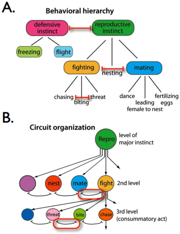
    1. Tinbergen N. The study of instinct. New York, NY: Clarendon Press/Oxford University Press; 1951.
    1. Miczek KA, de Almeida RM, Kravitz EA, Rissman EF, de Boer SF, Raine A. Neurobiology of escalated aggression and violence. J Neurosci. 2007;27:11803–11806. - PMC - PubMed
    1. LeDoux JE. Emotion circuits in the brain. Annu Rev Neurosci. 2000;23:155–184. - PubMed
    1. Paré D, Quirk GJ, LeDoux JE. New vistas on amygdala networks in conditioned fear. J Neurophysiol. 2004;92:1–9. - PubMed
    1. Ehrlich I, Humeau Y, Grenier F, Ciocchi S, Herry C, Luthi A. Amygdala inhibitory circuits and the control of fear memory. Neuron. 2009;62:757–771. - PubMed

Publication types