

Quantitative measurement of cerebral physiology using respiratory-calibrated MRI
- PMID: 22209811
- PMCID: PMC7100043
- DOI: 10.1016/j.neuroimage.2011.12.017
Quantitative measurement of cerebral physiology using respiratory-calibrated MRI
Abstract
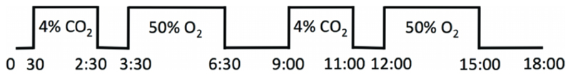
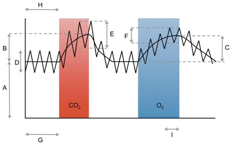
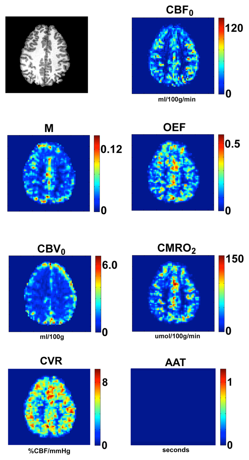
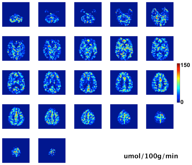
Functional magnetic resonance imaging typically measures signal increases arising from changes in the transverse relaxation rate over small regions of the brain and associates these with local changes in cerebral blood flow, blood volume and oxygen metabolism. Recent developments in pulse sequences and image analysis methods have improved the specificity of the measurements by focussing on changes in blood flow or changes in blood volume alone. However, FMRI is still unable to match the physiological information obtainable from positron emission tomography (PET), which is capable of quantitative measurements of blood flow and volume, and can indirectly measure resting metabolism. The disadvantages of PET are its cost, its availability, its poor spatial resolution and its use of ionising radiation. The MRI techniques introduced here address some of these limitations and provide physiological data comparable with PET measurements. We present an 18-minute MRI protocol that produces multi-slice whole-brain coverage and yields quantitative images of resting cerebral blood flow, cerebral blood volume, oxygen extraction fraction, CMRO(2), arterial arrival time and cerebrovascular reactivity of the human brain in the absence of any specific functional task. The technique uses a combined hyperoxia and hypercapnia paradigm with a modified arterial spin labelling sequence.
Copyright © 2011 Elsevier Inc. All rights reserved.
Figures
References
-
- Bulte DP, Chiarelli PA, Wise R, Jezzard P. Measurement of Cerebral Blood Volume in Humans Using Hyperoxic MRI Contrast. Journal of Magnetic Resonance Imaging. 2007a;26:894–899. - PubMed
MeSH terms
Substances
Grants and funding
LinkOut - more resources
Full Text Sources
Medical
