Review
. 2012 Mar;31(2):121-35.
doi: 10.1016/j.preteyeres.2011.12.001. Epub 2011 Dec 22.

The bisretinoids of retinal pigment epithelium

Affiliations
Review

The bisretinoids of retinal pigment epithelium

Janet R Sparrow et al. Prog Retin Eye Res. 2012 Mar.

Abstract

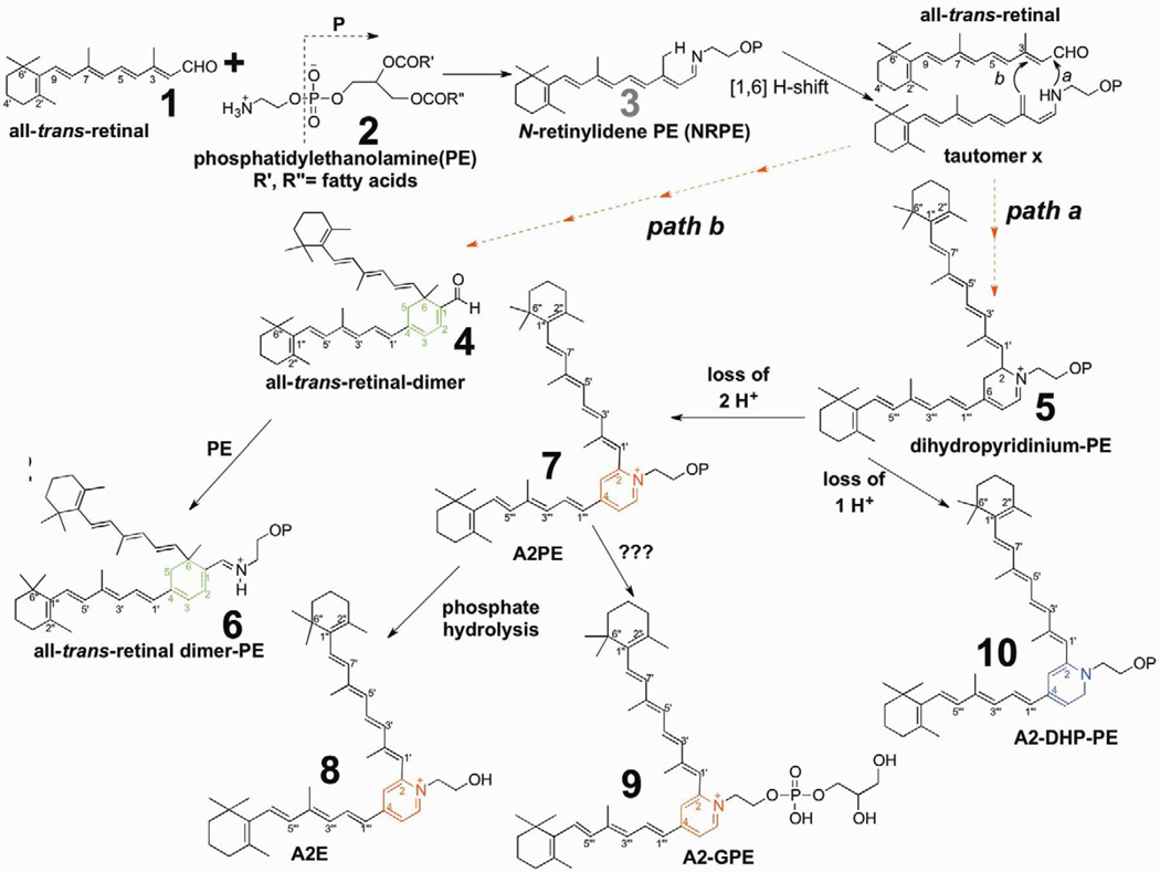
The retina exhibits an inherent autofluorescence that is imaged ophthalmoscopically as fundus autofluorescence. In clinical settings, fundus autofluorescence examination aids in the diagnosis and follow-up of many retinal disorders. Fundus autofluorescence originates from the complex mixture of bisretinoid fluorophores that are amassed by retinal pigment epithelial (RPE) cells as lipofuscin. Unlike the lipofuscin found in other cell-types, this material does not form as a result of oxidative stress. Rather, the formation is attributable to non-enzymatic reactions of vitamin A aldehyde in photoreceptor cells; transfer to RPE occurs upon phagocytosis of photoreceptor outer segments. These fluorescent pigments accumulate even in healthy photoreceptor cells and are generated as a consequence of the light capturing function of the cells. Nevertheless, the formation of this material is accelerated in some retinal disorders including recessive Stargardt disease and ELOVL4-related retinal degeneration. As such, these bisretinoid side-products are implicated in the disease processes that threaten vision. In this article, we review our current understanding of the composition of RPE lipofuscin, the structural characteristics of the various bisretinoids, their related spectroscopic features and the biosynthetic pathways by which they form. We will revisit factors known to influence the extent of the accumulation and therapeutic strategies being used to limit bisretinoid formation. Given their origin from vitamin A aldehyde, an isomer of the visual pigment chromophore, it is not surprising that the bisretinoids of retina are light sensitive molecules. Accordingly, we will discuss recent findings that implicate the photodegradation of bisretinoid in the etiology of age-related macular degeneration.


Figures

Figure 1
Figure 1
Proposed biosynthetic schemes for bisretinoid formation. All-trans-retinal that is released from opsin after photoisomerization of ground state 11-cis-retinal reacts with phosphatidylethanolamine (PE) in the disk membrane to produce the N-retinyl-phosphatidylethanolamine Schiff base (NRPE). NRPE undergoes a [1,6] H-shift producing tautomer X. Reaction with a second molecule of all-trans-retinal leads to path a and the formation of a bisretinoid phosphatidyl-dihydropyridinium molecule (dihydropyridinium-A2PE). Dihydropyridinium-A2PE automatically loses one hydrogen to produce A2-dihydropyridine-phosphatidylethanolamine (A2-DHP-PE) or it can eliminate 2 hydrogens to form A2PE, a phosphatidyl-pyridinium bis-retinoid. Hydrolysis of the phosphate ester of A2PE, probably by the lysosomal enzyme phospholipase D, yields A2E. Alternatively, all-trans-retinal can add to tautomer × and after ring closure all-trans-retinal dimer will form (path b). Subsequent Schiff base reaction with PE yields all-trans-retinal dimer-PE. Dashed arrows indicate multiple steps in the pathway. Note that OP indicates retention of phosphatidic acid (glycerol, phosphate and fatty acids) originating from PE.
Figure. 2
Figure. 2
A2E accumulates in central and peripheral retina of adult human eye. RPE/choroid samples obtained from human eye using 4 mm trephine. A. RPE/choroid from central (centered on fovea) retina. Tissue was analyzed by ultra performance liquid chromatography/mass spectrometry (MS) analysis (electrospray ion multi-mode ionization, ESI). Upper trace, chromatogram with absorbance monitoring at 430 nm. Inset, absorbance spectra of A2E and isoA2E. Lower trace, ESI-MS at m/z 592, the molecular weight of A2E. B. RPE/choroid from each of four retinal quadrants. Shown are absorbance spectra and molecular weights (m/z) for A2E and isoA2E obtained by analysis of samples from each quadrant. C. Chloroform/methanol extract of human RPE/choroid showing water soluble and chloroform/methanol soluble phases.
Figure 3
Figure 3
Ultra performance liquid chromatography/mass spectrometry analysis (electrospray ion multi-mode ionization) of eyes obtained from Abca4−/− mice (2–3 months of age, 8 eyes) with detection of N-retinylidene-PE (NRPE), the Schiff base adduct of all-trans-retinal and phosphatidylethanolamine. UPLC operated with BEH phenyl™ C18 reversed phase column with a mobile phase of acetonitrile/methanol (1:1) in water with 0.1% formic acid. Monitoring of 430 nm absorbance, fluorescence and molecular weight (m/z). NRPE is detected as multiple peaks reflecting differences in length of fatty acid chains and in unsaturation. The derived m/z values correspond to the lipid moieties indicated in parentheses. Insets above, UV-visible absorbance spectra of indicated eluting compounds. Structures, NRPE and plasmalogen-NRPE. NRPE has the usual ester bonds at the carbon 1 (sn-1) and carbon 2 (sn-2) positions of glycerol. In plasmalogen-NRPE the first position of the glycerol backbone is bonded to a vinyl-ether moiety (i.e. a carbon-carbon double bond on one side of a ether (C-O-C) linkage).
Figure 4
Figure 4
Detection of the bisretinoid A2PE in isolated bovine photoreceptor outer segments (POS). Ultra performance liquid chromatography/mass spectrometry analysis (electrospray ion multi-mode ionization) as in Fig. 2. A and B. In POS extract, a series of chromatographic peaks (A) exhibit m/z 1322.9 (B). UV-visible absorbances (Insets in A) are consistent with A2PE; the multiple peaks with same m/z are indicative of A2PE isomers. C. Incubation of POS with all-trans-retinal accentuates peak height (peaks 1–3 in A are increased in C), indicating that peaks 1–3 reflect compounds forming by reaction of all-trans-retinal. D and E. The fatty acid species in the A2PE detected in POS (A) are identified as stearic acid (18:0) and docosahexaenoic acid (DHA, 22:6) since A2PE synthesized from 1-stearoyl-2-docosahexaenoyl-sn-glycero-3-phosphoethanolamine has the same retention time (D) and m/z (E) as the compound in A and B.
Figure 5
Figure 5
A2PE and all-trans-retinal dimer synthesis: modulating factors. Reaction mixtures of all-trans-retinal and phosphatidylethanolamine (PE). Presented as equivalent ratios. A. A2PE formation when concentration of all-trans-retinal is varied. B. A2PE formation when concentration of PE is varied. C. All-trans-retinal dimer synthesis with varying PE concentration. D. A2PE yield using egg yolk-PE versus docosahexaenoic-PE (DHA-PE) as precursor. E. A2PE yield with varying levels of egg-PE and in the presence/absence of free DHA fatty acid (8 equivalents). F. A2PE yield is facilitated in presence of triethylamine; all-trans-retinal:PE ratio was 4:2. Synthetic reaction mixtures were prepared in chloroform/methanol (3:1) at 37°C. G. Detection of NRPE, all-trans-retinal dimer (atRAL-dimer) and A2PE by HPLC analysis (C4 column). Quantitation by integrating chromatographic peak areas. DHA-PE, 1,2-didocosahexaenoyl-sn-glycero-3-phosphoethanolamine. Major fatty acids in egg yolk-PE are 16:0, 18:0, 18:1, 18:2, 20:4 and 22:6.
Figure 6
Figure 6
Fluorescence emission spectra of RPE lipofuscin and cell-based A2E. Data recorded using confocal laser scanning fluorescence microscope (Nikon A1R MP) with a 60X objective in 6–nm increments while exciting with lasers at 488 and 561 nm. A. Emission spectra obtained from RPE monolayer (posterior) in cryostat sections of human eye (age 45 years) (field size 512 × 128 pixels, 0.21 um per pixel) and from A2E that had accumulated in ARPE-19 cells (field size 512 × 512 pixels, 0.42 microns per pixel). Emission data adjusted for laser power and pixel size. B. Emission spectra obtained from cryostat sections of eyes obtained from Abca4 null mutant mice at ages indicated in months (m). Field size was 512 × 64 pixels, 0.09 microns per pixel. Spectra were adjusted for pixel size and laser power. C. Fluorescence emission of A2E (in DMSO/buffered saline) when not irradiated (red trace) and when irradiated for the times indicated (blue traces). Emission peak wavelengths are indicated adjacent to each trace. Note the decrease in peak height and the peak shift to shorter wavelengths with increasing duration of irradiation. D. Emission spectra of RPE bisretinoid A2-GPE (diretinal glycerophosphethanolamine). Note red-shift with increasing excitation wavelength.
Figure 7
Figure 7
Photooxidation and photodegradation of all-trans-retinal dimer. Analysis by electrospray ion multi-mode ionization (ESI). Samples of all-trans-retinal were unirradiated (A) or irradiated (B) at 430 nm (hv). The series of peaks from m/z 551–679 reflect photooxidation at carbon-carbon double bonds in all-trans-retinal dimer. Lower mass peaks (< m/z 592) correspond to all-trans-retinal dimer photodegradation products. C. Proposed structures of the largest of the photodegradation products. Upper left, all-trans-retinal dimer exhibits absorbance peaks at 432 nm and 292 nm that can be assigned to long and short arms of the molecule, respectively.
Figure 8
Figure 8
Glyceraldehyde 3-phosphate dehydrogenase (GAPDH) activity is decreased in cells that have accumulated A2E and are irradiated at 430 nm (1.38 mW/cm2). ARPE-19 cells accumulate A2E into the lysosomal compartment. With this experimental model, the incidence of cell death is not elevated in A2E-containing cells versus untreated cells.
Figure 9
Figure 9
Photodegradation of the RPE bisretinoids A2E and all-trans-retinal dimer (atRAL dimer) releases the dicerbonyls methylglyoxal and glyoxal. Potential cleavage sites at carbon-carbon double bonds are presented (dotted lines) Depending on the photodegradation patterns, each molecule of A2E and all-trans-retinal dimer could release 2–3 molecules of methylglyoxal or glyoxal.
Figure 10
Figure 10
Senescence accelerated mice. These mice carry the Leu450 variant in Rpe65. A. Quantification of outer nuclear layer (ONL) thickness in senescence-accelerated resistant (SAMR) and senescence-accelerated prone (SAMP8) mice at age 6 month. Measurements are plotted as a function of distance from the optic nerve head (ONH) in the inferior and superior hemispheres. Values are mean ± SEM of 4 eyes. B,C. Light micrographs of SAMR and SAMP8 central retinas, 6 months of age. RPE is not included in the micrographs. D. HPLC measurements of A2E and isoA2E in eyecups of SAMR and SAMP8 mice. Values are mean ± SEM of 2–7 samples, 6 eyes per sample.

References

    1. Agbaga MP, Brush RS, Mandal MN, Henry K, Elliott MH, Anderson RE. Role of Stargardt-3 macular dystrophy protein (ELOVL4) in the biosynthesis of very long chain fatty acids. Proc Natl Acad Sci U S A. 2008;105:12843–12848. - PMC - PubMed
    1. Allikmets R, Singh N, Sun H, Shroyer NF, Hutchinson A, Chidambaram A, Gerrard B, Baird L, Stauffer D, Peiffer A, Rattner A, Smallwood P, Li Y, Anderson KL, Lewis RA, Nathans J, Leppert M, Dean M, Lupski JR. A photoreceptor cell-specific ATP-binding transporter gene (ABCR) is mutated in recessive Stargardt macular dystrophy. Nat Genet. 1997;15:236–246. - PubMed
    1. Allocca M, Doria M, Petrillo M, Colella P, Garcia-Hoyos M, Gibbs D, Kim SR, Maguire AM, Rex TS, Di Vicino U, Cutillo L, Sparrow JR, Williams DS, Bennett J, Auricchio A. Serotype-dependent packaging of large genes in adeno-associated viral vectors results in effective gene delivery in mice. J Clin Invest. 2008;118:1955–1964. - PMC - PubMed
    1. Anderson RE, Maude MB. Phospholipids of bovine outer segments. Biochemistry. 1970;9:3624–3628. - PubMed
    1. Beharry S, Zhong M, Molday RS. N-retinylidene-phosphatidylethanolamine is the preferred retinoid substrate for the photoreceptor-specific ABC transporter ABCA4 (ABCR) J Biol Chem. 2004;279:53972–53979. - PubMed

Publication types