. 2012 Mar;158(3):1426-38.
doi: 10.1104/pp.111.192385. Epub 2011 Dec 30.

Arabidopsis VILLIN2 and VILLIN3 are required for the generation of thick actin filament bundles and for directional organ growth

Affiliations

Arabidopsis VILLIN2 and VILLIN3 are required for the generation of thick actin filament bundles and for directional organ growth

Hannie S van der Honing et al. Plant Physiol. 2012 Mar.

Abstract

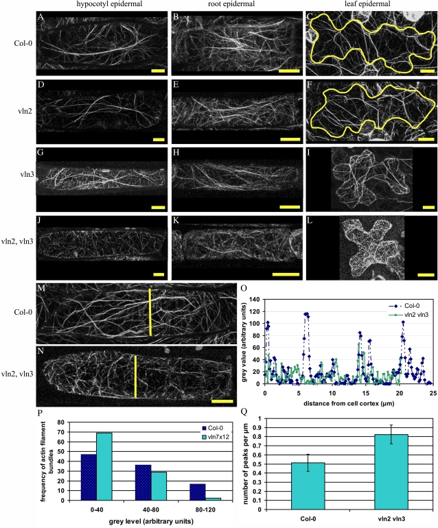
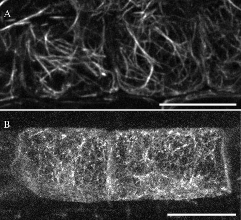
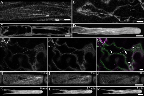
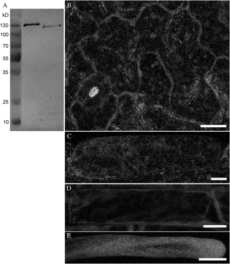
In plant cells, actin filament bundles serve as tracks for myosin-dependent organelle movement and play a role in the organization of the cytoplasm. Although virtually all plant cells contain actin filament bundles, the role of the different actin-bundling proteins remains largely unknown. In this study, we investigated the role of the actin-bundling protein villin in Arabidopsis (Arabidopsis thaliana). We used Arabidopsis T-DNA insertion lines to generate a double mutant in which VILLIN2 (VLN2) and VLN3 transcripts are truncated. Leaves, stems, siliques, and roots of vln2 vln3 double mutant plants are twisted, which is caused by local differences in cell length. Microscopy analysis of the actin cytoskeleton showed that in these double mutant plants, thin actin filament bundles are more abundant while thick actin filament bundles are virtually absent. In contrast to full-length VLN3, truncated VLN3 lacking the headpiece region does not rescue the phenotype of the vln2 vln3 double mutant. Our results show that villin is involved in the generation of thick actin filament bundles in several cell types and suggest that these bundles are involved in the regulation of coordinated cell expansion.


Figures

Figure 1.
Figure 1.
Characterization of the Arabidopsis villin gene family and T-DNA insertions in vln2 and vln3. A, Cladogram of the Arabidopsis villins, based on cDNA sequences. B, Locations of T-DNA inserts in vln2 and vln3. Gray boxes represent exons, and horizontal lines represent introns. T-DNA inserts (arrowheads) are not drawn to scale. C, Domain structure of villin. Arrowheads show locations corresponding to the locations of T-DNA inserts of vln2 and vln3. D, T-DNA insertions result in truncated transcripts in vln2, vln3, and vln2 vln3. Products could be amplified using a cDNA template using VLN-specific primers before the inserts, but when both primers (vln2) or the reverse primer (vln3) were designated for coding regions after the insert (Supplemental Fig. S3; Supplemental Table S1), products could not be amplified. Az, Azygous. E, A protein gel blot of Col-0, vln2, vln3, and vln2 vln3 root extracts probed with lily anti-villin antibody (Tominaga et al., 2000) shows that vln3 and vln2 vln3 do not contain (a truncated version of) the VLN3 protein.
Figure 2.
Figure 2.
Phenotype of vln2 vln3. A, Root morphology of azygous, (Az), vln2, vln3, and vln2 vln3 plants. Roots of both single mutants have the same appearance as azygous roots, but roots of double mutants grow in a curly, wavy manner. B, Phenotypes of 2-week-old plants. Leaves of the vln2 vln3 double mutant are twisted, but in single mutants, this twisting is absent. C, Phenotypes of 5-week-old plants. Branches of single mutants grow straight, similar to those of azygous plants, but in the double mutants, branches are curly and even show complete twists (arrow). This twisting also occurs in the fruit stalks. D, Twisting of double mutant branches and fruit stalks shown at a higher magnification. E, The rotational movements (circumnutation) of vln2 vln3 inflorescences have larger amplitudes than those of Col-0 inflorescences and are less regular (Supplemental Movie S1). [See online article for color version of this figure.]
Figure 3.
Figure 3.
Quantification of the vln2 vln3 phenotype. A, A total of 41% (n = 17) of the tops of inflorescence meristems of vln2 vln3 grow downward, while this never occurs in Col-0 (n = 22) and single mutant (n = 19 for vln2 and 17 for vln3) plants. B, The angle of siliques with respect to the plant axis of the vln2 vln3 double mutant is less regular than that of Col-0 and single mutant plants: siliques of vln2 vln3 grow in all directions at similar frequencies, while those of Col-0 and single mutant plants preferentially grow upward at an oblique angle. C and D, Leaf pavement (C; n = 26 for Col-0 and 61 for vln2 vln3) and fruit stalk epidermal (D; n = 68 for azygous and 67 for vln2 vln3) cell dimensions of vln2 vln3 are similar (Student’s t test, P > 0.05) to those of Col-0 plants, except for fruit stalk epidermal cell width, which is significantly higher (Student’s t test, P = 0.01) to that in vln2 vln3. Circularity reflects the ratio of cell area to cell perimeter and is defined as 4π area/perimeter2 (Vidali et al., 2007). Error bars in C and D represent sd. [See online article for color version of this figure.]
Figure 4.
Figure 4.
Thick actin bundles are absent in vln2 vln3, but thin bundles of actin filaments are more prominent. A to L, The actin organization (visualized with GFP:FABD2) in cells of both single mutants (D–I) is similar to that in Col-0 cells (A–C): thick bundles of actin filaments alternate with a more complex network of thin (bundles of) actin filaments. In the double mutant (J–L), thick actin filament bundles appear to be absent, while thin actin filament bundles seem more prominent. M to O, Representative intensity profiles of fluorescence intensity in a Col-0 (M) and a vln2 vln3 (N) fully elongated hypocotyl cell. High peaks represent thick actin filament bundles, while lower peaks represent thinner bundles. The yellow lines in M and N show the locations of the intensity profile in O. P, Frequency distribution of peaks belonging to three fluorescence intensity classes (determined for six cells for each genotype) in Col-0 and vln2 vln3. In Col-0 cells, peaks with a fluorescence intensity of 80 to 120 (representing thick actin filament bundles) are more abundant than in vln2 vln3, while peaks with a fluorescence intensity of 0 to 40 (representing thin[ner] actin filament bundles) are more abundant in vln2 vln3. Q, The number of peaks per micrometer shown for Col-0 and vln2 vln3. vln2 vln3 cells contain significantly more (Student’s t test, P = 0.04) actin filament bundles than Col-0 cells. Bars in A to N = 10 μm. Error bars in Q represent se. [See online article for color version of this figure.]
Figure 5.
Figure 5.
The irregular organ growth phenotype correlates with defects in actin organization in expanding cells. The actin organization in elongating root epidermal cells of the vln2 vln3 mutant (B) is disrupted when compared with the actin organization of Col-0 cells (A). The defects in actin organization resemble those in fully grown cells. The actin organization is visualized by GFP-FABD2 expression. Bars = 20 μm.
Figure 6.
Figure 6.
GFP:VLN3, expressed under the control of the VLN3 promoter, localizes to (bundles of) actin filaments. A to D, Representative images of complemented vln2 vln3 plants show that besides a cytoplasmic localization, GFP:VLN3 decorates (bundles of) actin filaments in hypocotyl epidermal (A), leaf epidermal (B), and root epidermal (C and D) cells. These images show fully grown hypocotyl epidermal (A) and leaf epidermal (B) cells and elongating root epidermal cells (C and D). In growing root hairs (D), GFP:VLN3 localizes to the long actin filament bundles oriented longitudinally to the cell’s long axis in the root hair tube. E to G, Coexpression of PVLN3:GFP:VLN3 and P35S:mCherry:FABD2 in tobacco demonstrates that GFP:VLN3 (E) and mCherry:FABD2 (F) colocalize (arrows) in leaf epidermal cells, although GFP:VLN3 does not localize to all actin filaments (arrowheads). G shows an overlay of E and F (GFP:VLN3, green; mCerry:FABD2, magenta). Image sequences of elongating root epidermal cells (H–J and K–M; root hairs) of complemented vln2 vln3 plants show that GFP:VLN3 localizes to (bundles of) actin filaments that reorganize over time. See also Supplemental Movies S2 and S3. Bars = 10 μm. [See online article for color version of this figure.]
Figure 7.
Figure 7.
GFP:VLN3ΔHP, expressed under the control of the VLN3 promoter, shows a cytoplasmic localization. A, Western blotting with an anti-GFP antibody reveals that GFP:VLN3-expressing plants express a fusion protein of approximately 137 kD, the expected mass of GFP:VLN3. GFP:VLN3ΔHP-expressing plants express a fusion protein of approximately 120 kD, which is the expected mass of GFP:VLN3ΔHP. B to E, Representative images of leaf epidermal (B), hypocotyl epidermal (C), and root epidermal (D) cells and a root hair (E) of vln2 vln3 plants in which GFP:VLN3ΔHP is expressed. In these plants, which are not rescued, GFP:VLN3ΔHP fluorescence is equally distributed throughout the cytoplasm. Bars = 10 μm.

Similar articles

Cited by

References

    1. Bartles JR. (2000) Parallel actin bundles and their multiple actin-bundling proteins. Curr Opin Cell Biol 12: 72–78 - PMC - PubMed
    1. Bouwmeester K, de Sain M, Weide R, Gouget A, Klamer S, Canut H, Govers F. (2011) The lectin receptor kinase LecRK-I.9 is a novel Phytophthora resistance component and a potential host target for a RXLR effector. PLoS Pathog 7: e1001327. - PMC - PubMed
    1. Brembu T, Winge P, Seem M, Bones AM. (2004) NAPP and PIRP encode subunits of a putative wave regulatory protein complex involved in plant cell morphogenesis. Plant Cell 16: 2335–2349 - PMC - PubMed
    1. Bretscher A, Weber K. (1980) Villin is a major protein of the microvillus cytoskeleton which binds both G and F actin in a calcium-dependent manner. Cell 20: 839–847 - PubMed
    1. Bryan J, Kurth MC. (1984) Actin-gelsolin interactions: evidence for two actin-binding sites. J Biol Chem 259: 7480–7487 - PubMed

MeSH terms

Substances